 Movie
Movie Controller

[English] 日本語
 Yorodumi
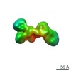
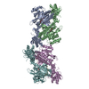
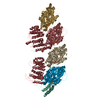
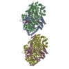
- PDB-7lbg: CryoEM structure of the HCMV Trimer gHgLgO in complex with human ... -
+ Open data

- Basic information
| Entry | Database: PDB / ID: 7lbg | ||||||
|---|---|---|---|---|---|---|---|
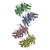
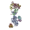
| Title | CryoEM structure of the HCMV Trimer gHgLgO in complex with human Transforming growth factor beta receptor type 3 and neutralizing fabs 13H11 and MSL-109 | ||||||
 Components Components |
| ||||||
 Keywords Keywords | VIRAL PROTEIN/Immune System / virus / receptor / complex / neutralizing antibody / VIRAL PROTEIN / VIRAL PROTEIN-Immune System complex | ||||||
| Function / homology |  Function and homology information Function and homology informationresponse to luteinizing hormone / epicardium-derived cardiac fibroblast cell development / transforming growth factor beta receptor activity, type III / transforming growth factor beta receptor complex assembly / inhibin-betaglycan-ActRII complex / TGFBR3 regulates activin signaling / response to follicle-stimulating hormone / muscular septum morphogenesis / definitive erythrocyte differentiation / TGFBR3 regulates FGF2 signaling ...response to luteinizing hormone / epicardium-derived cardiac fibroblast cell development / transforming growth factor beta receptor activity, type III / transforming growth factor beta receptor complex assembly / inhibin-betaglycan-ActRII complex / TGFBR3 regulates activin signaling / response to follicle-stimulating hormone / muscular septum morphogenesis / definitive erythrocyte differentiation / TGFBR3 regulates FGF2 signaling / vasculogenesis involved in coronary vascular morphogenesis / BMP binding / transforming growth factor beta receptor activity / regulation of transforming growth factor beta receptor signaling pathway / TGFBR3 PTM regulation / secondary palate development / ventricular compact myocardium morphogenesis / cardiac epithelial to mesenchymal transition / heart trabecula morphogenesis / TGFBR3 regulates TGF-beta signaling / type II transforming growth factor beta receptor binding / FGFR1b ligand binding and activation / activin binding / FGFR1c ligand binding and activation / transforming growth factor beta receptor binding / Signaling by BMP / Signaling by Activin / glycosaminoglycan binding / definitive hemopoiesis / heart trabecula formation / transforming growth factor beta binding / TGFBR3 expression / negative regulation of extracellular matrix assembly / positive regulation of cell migration involved in sprouting angiogenesis / ventricular septum morphogenesis / ventricular cardiac muscle tissue morphogenesis / positive regulation of transforming growth factor beta receptor signaling pathway / negative regulation of SMAD protein signal transduction / SMAD binding / outflow tract morphogenesis / TGF-beta receptor signaling activates SMADs / animal organ regeneration / positive regulation of SMAD protein signal transduction / fibroblast growth factor binding / epithelial to mesenchymal transition / cardiac muscle cell proliferation / BMP signaling pathway / positive regulation of cardiac muscle cell proliferation / heart morphogenesis / coreceptor activity / transforming growth factor beta receptor signaling pathway / negative regulation of cell migration / host cell endosome membrane / liver development / negative regulation of canonical NF-kappaB signal transduction / HCMV Late Events / PDZ domain binding / negative regulation of transforming growth factor beta receptor signaling pathway / response to prostaglandin E / negative regulation of epithelial cell proliferation / HCMV Early Events / transmembrane signaling receptor activity / cell migration / heparin binding / basolateral plasma membrane / host cell Golgi apparatus / entry receptor-mediated virion attachment to host cell / response to hypoxia / signaling receptor complex / intracellular signal transduction / immune response / fusion of virus membrane with host plasma membrane / external side of plasma membrane / viral envelope / positive regulation of gene expression / symbiont entry into host cell / host cell plasma membrane / virion membrane / cell surface / : / extracellular exosome / extracellular region / plasma membrane / cytosol Similarity search - Function | ||||||
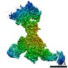
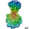
| Biological species |   Human cytomegalovirus Human cytomegalovirus Homo sapiens (human) Homo sapiens (human) | ||||||
| Method | ELECTRON MICROSCOPY / single particle reconstruction / cryo EM / Resolution: 2.6 Å | ||||||
 Authors Authors | Kschonsak, M. / Rouge, L. / Arthur, C.P. / Hoangdung, H. / Patel, N. / Kim, I. / Johnson, M. / Kraft, E. / Rohou, A.L. / Gill, A. ...Kschonsak, M. / Rouge, L. / Arthur, C.P. / Hoangdung, H. / Patel, N. / Kim, I. / Johnson, M. / Kraft, E. / Rohou, A.L. / Gill, A. / Martinez-Martin, N. / Payandeh, J. / Ciferri, C. | ||||||
 Citation Citation | Journal: Cell / Year: 2021 Title: Structures of HCMV Trimer reveal the basis for receptor recognition and cell entry. Authors: Marc Kschonsak / Lionel Rougé / Christopher P Arthur / Ho Hoangdung / Nidhi Patel / Ingrid Kim / Matthew C Johnson / Edward Kraft / Alexis L Rohou / Avinash Gill / Nadia Martinez-Martin / ...Authors: Marc Kschonsak / Lionel Rougé / Christopher P Arthur / Ho Hoangdung / Nidhi Patel / Ingrid Kim / Matthew C Johnson / Edward Kraft / Alexis L Rohou / Avinash Gill / Nadia Martinez-Martin / Jian Payandeh / Claudio Ciferri /  Abstract: Human cytomegalovirus (HCMV) infects the majority of the human population and represents the leading viral cause of congenital birth defects. HCMV utilizes the glycoproteins gHgLgO (Trimer) to bind ...Human cytomegalovirus (HCMV) infects the majority of the human population and represents the leading viral cause of congenital birth defects. HCMV utilizes the glycoproteins gHgLgO (Trimer) to bind to platelet-derived growth factor receptor alpha (PDGFRα) and transforming growth factor beta receptor 3 (TGFβR3) to gain entry into multiple cell types. This complex is targeted by potent neutralizing antibodies and represents an important candidate for therapeutics against HCMV. Here, we determine three cryogenic electron microscopy (cryo-EM) structures of the trimer and the details of its interactions with four binding partners: the receptor proteins PDGFRα and TGFβR3 as well as two broadly neutralizing antibodies. Trimer binding to PDGFRα and TGFβR3 is mutually exclusive, suggesting that they function as independent entry receptors. In addition, Trimer-PDGFRα interaction has an inhibitory effect on PDGFRα signaling. Our results provide a framework for understanding HCMV receptor engagement, neutralization, and the development of anti-viral strategies against HCMV. | ||||||
| History |
|
- Structure visualization
| Movie |
Movie viewer |
|---|---|
| Structure viewer | Molecule: MolmilJmol/JSmol |
- Downloads & links
-Download
| PDBx/mmCIF format | 7lbg.cif.gz | 362.2 KB | Display | PDBx/mmCIF format |
|---|---|---|---|---|
| PDB format | pdb7lbg.ent.gz | 274.3 KB | Display | PDB format |
| PDBx/mmJSON format | 7lbg.json.gz | Tree view | PDBx/mmJSON format | |
| Others |  Other downloads Other downloads |
-Validation report
| Arichive directory | https://data.pdbj.org/pub/pdb/validation_reports/lb/7lbgftp://data.pdbj.org/pub/pdb/validation_reports/lb/7lbg | HTTPS FTP |
|---|
-Related structure data
| Related structure data |  23254MC 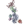 7lbeC 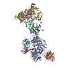 7lbfC M: map data used to model this data C: citing same article ( |
|---|---|
| Similar structure data |

-Links
 PDBj
PDBj


- Assembly
| Deposited unit | 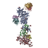
|
|---|---|
| 1 |
|
-Components
-Envelope glycoprotein ... , 3 types, 3 molecules ABC
| #1: Protein | Mass: 87311.273 Da / Num. of mol.: 1 Source method: isolated from a genetically manipulated source Source: (gene. exp.) Human cytomegalovirus (strain Merlin) / Strain: Merlin / Gene: gH, UL75 / Cell line (production host): Expi293 / Production host: Homo sapiens (human) / References: UniProt: Q6SW67 |
|---|---|
| #2: Protein | Mass: 30846.492 Da / Num. of mol.: 1 Source method: isolated from a genetically manipulated source Source: (gene. exp.) Human cytomegalovirus (strain Merlin) / Strain: Merlin / Gene: gL, UL115 / Cell line (production host): Expi293 / Production host: Homo sapiens (human) / References: UniProt: F5HCH8 |
| #3: Protein | Mass: 58298.504 Da / Num. of mol.: 1 Source method: isolated from a genetically manipulated source Source: (gene. exp.) Human cytomegalovirus / Gene: UL74 / Cell line (production host): Expi293 / Production host: Homo sapiens (human) / References: UniProt: Q8BCU3 |
-Protein , 1 types, 1 molecules D
| #4: Protein | Mass: 87362.203 Da / Num. of mol.: 1 Source method: isolated from a genetically manipulated source Source: (gene. exp.) Homo sapiens (human) / Gene: TGFBR3 / Production host:   Spodoptera frugiperda (fall armyworm) / References: UniProt: Q03167 Spodoptera frugiperda (fall armyworm) / References: UniProt: Q03167 |
|---|
-Antibody , 4 types, 4 molecules EFGH
| #5: Antibody | Mass: 25780.020 Da / Num. of mol.: 1 Source method: isolated from a genetically manipulated source Source: (gene. exp.) Homo sapiens (human) / Production host:  |
|---|---|
| #6: Antibody | Mass: 26600.086 Da / Num. of mol.: 1 Source method: isolated from a genetically manipulated source Source: (gene. exp.) Homo sapiens (human) / Production host: |
| #7: Antibody | Mass: 28355.809 Da / Num. of mol.: 1 Source method: isolated from a genetically manipulated source Source: (gene. exp.) Homo sapiens (human) / Production host: |
| #8: Antibody | Mass: 27547.818 Da / Num. of mol.: 1 Source method: isolated from a genetically manipulated source Source: (gene. exp.) Homo sapiens (human) / Production host: |
-Sugars , 3 types, 19 molecules 
| #9: Polysaccharide | 2-acetamido-2-deoxy-beta-D-glucopyranose-(1-4)-2-acetamido-2-deoxy-beta-D-glucopyranose |
|---|---|
| #10: Polysaccharide | alpha-D-mannopyranose-(1-2)-alpha-D-mannopyranose-(1-3)-[alpha-D-mannopyranose-(1-3)-alpha-D- ...alpha-D-mannopyranose-(1-2)-alpha-D-mannopyranose-(1-3)-[alpha-D-mannopyranose-(1-3)-alpha-D-mannopyranose-(1-6)]beta-D-mannopyranose-(1-4)-2-acetamido-2-deoxy-beta-D-glucopyranose-(1-4)-2-acetamido-2-deoxy-beta-D-glucopyranose Source method: isolated from a genetically manipulated source |
| #11: Sugar | ChemComp-NAG /  Type: D-saccharide, beta linking / Mass: 221.208 Da / Num. of mol.: 17 / Source method: obtained synthetically / Formula: C8H15NO6 Type: D-saccharide, beta linking / Mass: 221.208 Da / Num. of mol.: 17 / Source method: obtained synthetically / Formula: C8H15NO6 |
-Details
| Has ligand of interest | N |
|---|---|
| Has protein modification | Y |
-Experimental details
-Experiment
| Experiment | Method: ELECTRON MICROSCOPY |
|---|---|
| EM experiment | Aggregation state: 2D ARRAY / 3D reconstruction method: single particle reconstruction |
- Sample preparation
| Component |
| ||||||||||||||||||||||||
|---|---|---|---|---|---|---|---|---|---|---|---|---|---|---|---|---|---|---|---|---|---|---|---|---|---|
| Molecular weight | Value: 0.36 MDa / Experimental value: NO | ||||||||||||||||||||||||
| Source (natural) |
| ||||||||||||||||||||||||
| Source (recombinant) |
| ||||||||||||||||||||||||
| Buffer solution | pH: 7.5 | ||||||||||||||||||||||||
| Buffer component |
| ||||||||||||||||||||||||
| Specimen | Conc.: 0.5 mg/ml / Embedding applied: NO / Shadowing applied: NO / Staining applied: NO / Vitrification applied: YES / Details: This sample was monodisperse | ||||||||||||||||||||||||
| Specimen support | Grid material: GOLD / Grid mesh size: 300 divisions/in. / Grid type: Quantifoil R1.2/1.3 | ||||||||||||||||||||||||
| Vitrification | Instrument: LEICA EM GP / Cryogen name: ETHANE / Humidity: 100 % / Chamber temperature: 277 K / Details: blot for 3.5 seconds before plunging |
- Electron microscopy imaging
| Experimental equipment |  Model: Titan Krios / Image courtesy: FEI Company |
|---|---|
| Microscopy | Model: FEI TITAN KRIOS |
| Electron gun | Electron source:  FIELD EMISSION GUN / Accelerating voltage: 300 kV / Illumination mode: FLOOD BEAM FIELD EMISSION GUN / Accelerating voltage: 300 kV / Illumination mode: FLOOD BEAM |
| Electron lens | Mode: BRIGHT FIELD / Nominal magnification: 165000 X |
| Specimen holder | Cryogen: NITROGEN / Specimen holder model: FEI TITAN KRIOS AUTOGRID HOLDER |
| Image recording | Average exposure time: 10 sec. / Electron dose: 50 e/Å2 / Film or detector model: GATAN K2 SUMMIT (4k x 4k) / Num. of grids imaged: 1 / Num. of real images: 19993 / Details: Images were collected in 50 frames every 0.2 s |
| Image scans | Movie frames/image: 50 |
- Processing
| EM software |
| ||||||||||||||||||||||||||||||||||||||||||||||||||||||||||||
|---|---|---|---|---|---|---|---|---|---|---|---|---|---|---|---|---|---|---|---|---|---|---|---|---|---|---|---|---|---|---|---|---|---|---|---|---|---|---|---|---|---|---|---|---|---|---|---|---|---|---|---|---|---|---|---|---|---|---|---|---|---|
| CTF correction | Type: PHASE FLIPPING AND AMPLITUDE CORRECTION | ||||||||||||||||||||||||||||||||||||||||||||||||||||||||||||
| Particle selection | Num. of particles selected: 2780519 | ||||||||||||||||||||||||||||||||||||||||||||||||||||||||||||
| Symmetry | Point symmetry: C1 (asymmetric) | ||||||||||||||||||||||||||||||||||||||||||||||||||||||||||||
| 3D reconstruction | Resolution: 2.6 Å / Resolution method: FSC 0.143 CUT-OFF / Num. of particles: 2737199 Details: Used a score threshold of 0.25 for final 3D reconstruction. Map used for model refinements is a composite map after combining 2 focussed maps with PHENIX Symmetry type: POINT | ||||||||||||||||||||||||||||||||||||||||||||||||||||||||||||
| Atomic model building | Protocol: AB INITIO MODEL / Space: REAL |
