 Movie
Movie Controller


+ Open data

- Basic information
| Entry | Database: PDB / ID: 7lb6 | |||||||||
|---|---|---|---|---|---|---|---|---|---|---|
| Title | PDX1.2/PDX1.3 co-expression complex | |||||||||
 Components Components |
| |||||||||
 Keywords Keywords | PLANT PROTEIN / pseudoenzyme / dodecamer / vitamin B6 | |||||||||
| Function / homology |  Function and homology information Function and homology informationresponse to non-ionic osmotic stress / response to lipid hydroperoxide / chlorophyll metabolic process / amine-lyase activity / pyridoxal 5'-phosphate synthase (glutamine hydrolysing) / pyridoxal 5'-phosphate synthase (glutamine hydrolysing) activity / pyridoxal 5'-phosphate biosynthetic process / hyperosmotic salinity response / pyridoxine biosynthetic process / amino acid metabolic process ...response to non-ionic osmotic stress / response to lipid hydroperoxide / chlorophyll metabolic process / amine-lyase activity / pyridoxal 5'-phosphate synthase (glutamine hydrolysing) / pyridoxal 5'-phosphate synthase (glutamine hydrolysing) activity / pyridoxal 5'-phosphate biosynthetic process / hyperosmotic salinity response / pyridoxine biosynthetic process / amino acid metabolic process / response to UV-B / response to salt stress / endomembrane system / response to oxidative stress / protein heterodimerization activity / protein homodimerization activity / plasma membrane / cytosol Similarity search - Function | |||||||||
| Biological species |  | |||||||||
| Method | ELECTRON MICROSCOPY / single particle reconstruction / cryo EM / Resolution: 3.16 Å | |||||||||
 Authors Authors | Novikova, I.V. / Evans, J.E. | |||||||||
| Funding support |  United States, 2items United States, 2items
| |||||||||
 Citation Citation | Journal: ACS Chem Biol / Year: 2021 Title: Tunable Heteroassembly of a Plant Pseudoenzyme-Enzyme Complex. Authors: Irina V Novikova / Mowei Zhou / Chen Du / Marcelina Parra / Doo Nam Kim / Zachary L VanAernum / Jared B Shaw / Hanjo Hellmann / Vicki H Wysocki / James E Evans / Abstract: Pseudoenzymes have emerged as key regulatory elements in all kingdoms of life despite being catalytically nonactive. Yet many factors defining why one protein is active while its homologue is ...Pseudoenzymes have emerged as key regulatory elements in all kingdoms of life despite being catalytically nonactive. Yet many factors defining why one protein is active while its homologue is inactive remain uncertain. For pseudoenzyme-enzyme pairs, the similarity of both subunits can often hinder conventional characterization approaches. In plants, a pseudoenzyme, PDX1.2, positively regulates vitamin B production by association with its active catalytic homologues such as PDX1.3 through an unknown assembly mechanism. Here we used an integrative experimental approach to learn that such pseudoenzyme-enzyme pair associations result in heterocomplexes of variable stoichiometry, which are unexpectedly tunable. We also present the atomic structure of the PDX1.2 pseudoenzyme as well as the population averaged PDX1.2-PDX1.3 pseudoenzyme-enzyme pair. Finally, we dissected hetero-dodecamers of each stoichiometry to understand the arrangement of monomers in the heterocomplexes and identified symmetry-imposed preferences in PDX1.2-PDX1.3 interactions. Our results provide a new model of pseudoenzyme-enzyme interactions and their native heterogeneity. | |||||||||
| History |
|
- Structure visualization
| Movie |
Movie viewer |
|---|---|
| Structure viewer | Molecule: MolmilJmol/JSmol |
- Downloads & links
-Download
| PDBx/mmCIF format | 7lb6.cif.gz | 1 MB | Display | PDBx/mmCIF format |
|---|---|---|---|---|
| PDB format | pdb7lb6.ent.gz | 857.9 KB | Display | PDB format |
| PDBx/mmJSON format | 7lb6.json.gz | Tree view | PDBx/mmJSON format | |
| Others |  Other downloads Other downloads |
-Validation report
| Arichive directory | https://data.pdbj.org/pub/pdb/validation_reports/lb/7lb6ftp://data.pdbj.org/pub/pdb/validation_reports/lb/7lb6 | HTTPS FTP |
|---|
-Related structure data
| Related structure data | 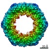 23264MC  7lb5C C: citing same article ( M: map data used to model this data |
|---|---|
| Similar structure data |


-Links
 PDBj
PDBj
- Assembly
| Deposited unit | 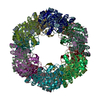
|
|---|---|
| 1 |
|
-Components
| #1: Protein | Mass: 37408.004 Da / Num. of mol.: 12 Source method: isolated from a genetically manipulated source Source: (gene. exp.) #2: Protein | Mass: 36475.531 Da / Num. of mol.: 12 Source method: isolated from a genetically manipulated source Source: (gene. exp.) References: UniProt: Q8L940, pyridoxal 5'-phosphate synthase (glutamine hydrolysing) |
|---|
-Experimental details
-Experiment
| Experiment | Method: ELECTRON MICROSCOPY |
|---|---|
| EM experiment | Aggregation state: PARTICLE / 3D reconstruction method: single particle reconstruction |
- Sample preparation
| Component | Name: PDX1.2/PDX1.3 heterododecamer / Type: COMPLEX Details: Represents an average 3D class for all PDX1.2/PDX1.3 heterododecamers Entity ID: all / Source: RECOMBINANT |
|---|---|
| Molecular weight | Value: 0.445 MDa / Experimental value: YES |
| Source (natural) | Organism: |
| Source (recombinant) | Organism: |
| Buffer solution | pH: 7.5 / Details: 50 mM Tris, 150 mM NaCl, 4 mM DTT |
| Specimen | Conc.: 0.2 mg/ml / Embedding applied: NO / Shadowing applied: NO / Staining applied: NO / Vitrification applied: YES |
| Specimen support | Details: at 15 mA / Grid material: GOLD / Grid mesh size: 300 divisions/in. / Grid type: Quantifoil R1.2/1.3 |
| Vitrification | Instrument: LEICA EM GP / Cryogen name: ETHANE / Humidity: 85 % / Chamber temperature: 298 K |
- Electron microscopy imaging
| Experimental equipment |  Model: Titan Krios / Image courtesy: FEI Company |
|---|---|
| Microscopy | Model: FEI TITAN KRIOS |
| Electron gun | Electron source:  FIELD EMISSION GUN / Accelerating voltage: 300 kV / Illumination mode: FLOOD BEAM FIELD EMISSION GUN / Accelerating voltage: 300 kV / Illumination mode: FLOOD BEAM |
| Electron lens | Mode: BRIGHT FIELD / Cs: 2.7 mm |
| Image recording | Electron dose: 100 e/Å2 / Detector mode: SUPER-RESOLUTION / Film or detector model: GATAN K2 SUMMIT (4k x 4k) |
- Processing
| EM software |
| |||||||||||||||||||||||||||||||||||||||||||||
|---|---|---|---|---|---|---|---|---|---|---|---|---|---|---|---|---|---|---|---|---|---|---|---|---|---|---|---|---|---|---|---|---|---|---|---|---|---|---|---|---|---|---|---|---|---|---|
| CTF correction | Type: PHASE FLIPPING AND AMPLITUDE CORRECTION | |||||||||||||||||||||||||||||||||||||||||||||
| Particle selection | Num. of particles selected: 510660 | |||||||||||||||||||||||||||||||||||||||||||||
| Symmetry | Point symmetry: D6 (2x6 fold dihedral) | |||||||||||||||||||||||||||||||||||||||||||||
| 3D reconstruction | Resolution: 3.16 Å / Resolution method: FSC 0.143 CUT-OFF / Num. of particles: 286642 / Symmetry type: POINT |
