 Movie
Movie Controller

[English] 日本語
 Yorodumi
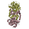
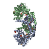
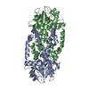
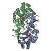
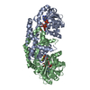
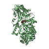
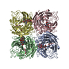
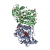
- PDB-7lat: Campylobacter jejuni keto-acid reductoisomerase in complex with Mg2+ -
+ Open data

- Basic information
| Entry | Database: PDB / ID: 7lat | ||||||
|---|---|---|---|---|---|---|---|
| Title | Campylobacter jejuni keto-acid reductoisomerase in complex with Mg2+ | ||||||
 Components Components | Ketol-acid reductoisomerase | ||||||
 Keywords Keywords | ISOMERASE / metalloenzyme / reductoisomerase | ||||||
| Function / homology |  Function and homology information Function and homology informationketol-acid reductoisomerase (NADP+) / ketol-acid reductoisomerase activity / L-valine biosynthetic process / : / isomerase activity / NADP binding / magnesium ion binding / cytosol Similarity search - Function | ||||||
| Biological species |   Campylobacter jejuni (Campylobacter) Campylobacter jejuni (Campylobacter) | ||||||
| Method |  X-RAY DIFFRACTION / SYNCHROTRON / MOLECULAR REPLACEMENT / Resolution: 2.47 Å X-RAY DIFFRACTION / SYNCHROTRON / MOLECULAR REPLACEMENT / Resolution: 2.47 Å | ||||||
 Authors Authors | Guddat, L.W. | ||||||
| Funding support |  Australia, 1items Australia, 1items
| ||||||
 Citation Citation | Journal: Acs Catalysis / Year: 2024 Title: Mapping of the Reaction Trajectory catalyzed by Class I Ketol-Acid Reductoisomerase Authors: Lin, X. / Lonhienne, T. / Lv, Y. / Kurz, J. / McGeary, R. / Schenk, G. / Guddat, L.W. | ||||||
| History |
|
- Structure visualization
| Structure viewer | Molecule: MolmilJmol/JSmol |
|---|
- Downloads & links
-Download
| PDBx/mmCIF format | 7lat.cif.gz | 94.2 KB | Display | PDBx/mmCIF format |
|---|---|---|---|---|
| PDB format | pdb7lat.ent.gz | 57.3 KB | Display | PDB format |
| PDBx/mmJSON format | 7lat.json.gz | Tree view | PDBx/mmJSON format | |
| Others |  Other downloads Other downloads |
-Validation report
| Arichive directory | https://data.pdbj.org/pub/pdb/validation_reports/la/7latftp://data.pdbj.org/pub/pdb/validation_reports/la/7lat | HTTPS FTP |
|---|
-Related structure data
| Related structure data | 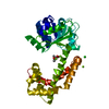 8swmC 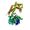 8sxdC  8upnC 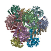 8uppC 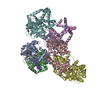 8upqC 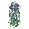 4ypoS S: Starting model for refinement C: citing same article ( |
|---|---|
| Similar structure data |


-Links
 PDBj
PDBj


- Assembly
| Deposited unit | 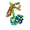
| ||||||||||||
|---|---|---|---|---|---|---|---|---|---|---|---|---|---|
| 1 | 
| ||||||||||||
| Unit cell |
| ||||||||||||
| Components on special symmetry positions |
|
-Components
| #1: Protein | Mass: 35868.223 Da / Num. of mol.: 1 Source method: isolated from a genetically manipulated source Source: (gene. exp.) Campylobacter jejuni (Campylobacter) / Gene: ilvC, CW563_00670 / Production host: | ||||
|---|---|---|---|---|---|
| #2: Chemical |   Mass: 24.305 Da / Num. of mol.: 2 / Source method: isolated from a natural source / Formula: Mg / Feature type: SUBJECT OF INVESTIGATION Mass: 24.305 Da / Num. of mol.: 2 / Source method: isolated from a natural source / Formula: Mg / Feature type: SUBJECT OF INVESTIGATION#3: Water | ChemComp-HOH / |  Mass: 18.015 Da / Num. of mol.: 105 / Source method: isolated from a natural source / Formula: H2O Mass: 18.015 Da / Num. of mol.: 105 / Source method: isolated from a natural source / Formula: H2OHas ligand of interest | Y | |
-Experimental details
-Experiment
| Experiment | Method: X-RAY DIFFRACTION / Number of used crystals: 1 |
|---|
- Sample preparation
| Crystal | Density Matthews: 2.53 Å3/Da / Density % sol: 51.38 % |
|---|---|
| Crystal grow | Temperature: 293 K / Method: vapor diffusion, hanging drop / pH: 5.5 Details: Ppt buffer 0.2 M Li2SO4, 0.1 M Bis-Tris pH 5.5, 20% PEG3350. The well solution (which was not added to the drop) 35% PEG3350, 250 mM MgCl2, 20 mM Tris-HCl pH 8.0. |
-Data collection
| Diffraction | Mean temperature: 100 K / Serial crystal experiment: N |
|---|---|
| Diffraction source | Source: SYNCHROTRON / Site: Australian Synchrotron / Beamline: MX1 / Wavelength: 1 Å |
| Detector | Type: ADSC QUANTUM 210r / Detector: CCD / Date: Jul 1, 2016 |
| Radiation | Monochromator: Double-crystal Si(111) / Protocol: SINGLE WAVELENGTH / Monochromatic (M) / Laue (L): M / Scattering type: x-ray |
| Radiation wavelength | Wavelength: 1 Å / Relative weight: 1 |
| Reflection | Resolution: 2.47→45.52 Å / Num. obs: 12844 / % possible obs: 99.9 % / Observed criterion σ(F): 0 / Observed criterion σ(I): 0 / Redundancy: 23.5 % / Biso Wilson estimate: 37.3319105008 Å2 / Rmerge(I) obs: 0.127 / Rpim(I) all: 0.027 / Net I/σ(I): 20.9 |
| Reflection shell | Resolution: 2.47→2.59 Å / Redundancy: 22.1 % / Rmerge(I) obs: 0.816 / Mean I/σ(I) obs: 3.5 / Num. unique obs: 1441 / Rpim(I) all: 0.178 / % possible all: 99 |
- Processing
| Software |
| ||||||||||||||||||||||||||||||||||||||||||||||||||||||||||||||||||||||
|---|---|---|---|---|---|---|---|---|---|---|---|---|---|---|---|---|---|---|---|---|---|---|---|---|---|---|---|---|---|---|---|---|---|---|---|---|---|---|---|---|---|---|---|---|---|---|---|---|---|---|---|---|---|---|---|---|---|---|---|---|---|---|---|---|---|---|---|---|---|---|---|
| Refinement | Method to determine structure: MOLECULAR REPLACEMENT Starting model: 4YPO Resolution: 2.47→45.52 Å / SU ML: 0.26725473031 / Cross valid method: FREE R-VALUE / σ(F): 1.36 / Phase error: 22.4152515085
| ||||||||||||||||||||||||||||||||||||||||||||||||||||||||||||||||||||||
| Solvent computation | Shrinkage radii: 0.9 Å / VDW probe radii: 1.11 Å / Solvent model: FLAT BULK SOLVENT MODEL | ||||||||||||||||||||||||||||||||||||||||||||||||||||||||||||||||||||||
| Displacement parameters | Biso mean: 40.3898217922 Å2 | ||||||||||||||||||||||||||||||||||||||||||||||||||||||||||||||||||||||
| Refinement step | Cycle: LAST / Resolution: 2.47→45.52 Å
| ||||||||||||||||||||||||||||||||||||||||||||||||||||||||||||||||||||||
| Refine LS restraints |
| ||||||||||||||||||||||||||||||||||||||||||||||||||||||||||||||||||||||
| LS refinement shell |
|
