 Movie
Movie Controller

[English] 日本語
 Yorodumi
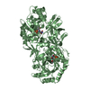
- PDB-2c54: gdp-mannose-3', 5' -epimerase (arabidopsis thaliana),k178r, with ... -
+ Open data

- Basic information
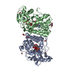
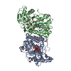
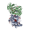
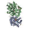
| Entry | Database: PDB / ID: 2c54 | ||||||
|---|---|---|---|---|---|---|---|
| Title | gdp-mannose-3', 5' -epimerase (arabidopsis thaliana),k178r, with gdp-beta-l-gulose and gdp-4-keto-beta-l-gulose bound in active site. | ||||||
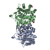
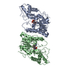
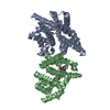
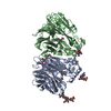
 Components Components | GDP-MANNOSE-3', 5'-EPIMERASE | ||||||
 Keywords Keywords | ISOMERASE / 3' 5'-EPIMERASE / SHORT CHAIN DEHYDRATASE/REDUCTASE / GDP-MANNOSE / GDP-GULOSE / GDP-GALACTOSE / KETO INTERMEDIATE / VITAMIN C / SDR / ASCORBATE BIOSYNTHESIS / NAD | ||||||
| Function / homology |  Function and homology information Function and homology informationGDP-mannose 3,5-epimerase / GDP-mannose 3,5-epimerase activity / L-ascorbic acid biosynthetic process / racemase and epimerase activity / plastid / NAD binding / cytoplasmic stress granule / cytosol Similarity search - Function | ||||||
| Biological species |  | ||||||
| Method |  X-RAY DIFFRACTION / SYNCHROTRON / MOLECULAR REPLACEMENT / Resolution: 1.5 Å X-RAY DIFFRACTION / SYNCHROTRON / MOLECULAR REPLACEMENT / Resolution: 1.5 Å | ||||||
 Authors Authors | Major, L.L. / Wolucka, B.A. / Naismith, J.H. | ||||||
 Citation Citation | Journal: J. Am. Chem. Soc. / Year: 2005 Title: Structure and function of GDP-mannose-3',5'-epimerase: an enzyme which performs three chemical reactions at the same active site. Authors: Major, L.L. / Wolucka, B.A. / Naismith, J.H. | ||||||
| History |
|
- Structure visualization
| Structure viewer | Molecule: MolmilJmol/JSmol |
|---|
- Downloads & links
-Download
| PDBx/mmCIF format | 2c54.cif.gz | 357.9 KB | Display | PDBx/mmCIF format |
|---|---|---|---|---|
| PDB format | pdb2c54.ent.gz | 290 KB | Display | PDB format |
| PDBx/mmJSON format | 2c54.json.gz | Tree view | PDBx/mmJSON format | |
| Others |  Other downloads Other downloads |
-Validation report
| Arichive directory | https://data.pdbj.org/pub/pdb/validation_reports/c5/2c54ftp://data.pdbj.org/pub/pdb/validation_reports/c5/2c54 | HTTPS FTP |
|---|
-Related structure data


-Links
 PDBj
PDBj- Assembly
| Deposited unit | 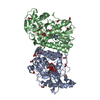
| ||||||||
|---|---|---|---|---|---|---|---|---|---|
| 1 |
| ||||||||
| Unit cell |
|
-Components
-Protein , 1 types, 2 molecules AB
| #1: Protein | Mass: 42971.613 Da / Num. of mol.: 2 / Mutation: YES Source method: isolated from a genetically manipulated source Details: GDP-BETA-L-GULOSE WAS REFINED USING GMP (DEFINED AS GDP, MODIFIED IN THE CIF FILE TO REMOVE THE SECOND PHOSPHATE GROUP) AND GULOSE-MONOPHOSPHATE (GUL), LINKING THE GDP O3A TO THE GUL PB. GDP- ...Details: GDP-BETA-L-GULOSE WAS REFINED USING GMP (DEFINED AS GDP, MODIFIED IN THE CIF FILE TO REMOVE THE SECOND PHOSPHATE GROUP) AND GULOSE-MONOPHOSPHATE (GUL), LINKING THE GDP O3A TO THE GUL PB. GDP-BETA-L-4-KETO-GULOSE WAS REFINED USING GMP (DEFINED AS GDP, MODIFIED IN THE CIF FILE TO REMOVE THE SECOND PHOSPHATE GROUP) AND 4-KETO-GULOSE- MONOPHOSPHATE (4KG), LINKING THE GDP O3A TO THE 4KG PB NAD WAS REFINED USING ADP AND A NICOTINAMIDE RING (NI3), LINKING THE ADP PB TO THE NI3 O5* NADH WAS REFINED USING ADP AND A SKEWED REDUCED NICOTINAMIDE RING (NDH), LINKING THE ADP PB TO THE NDH O5* Source: (gene. exp.)  |
|---|
-Non-polymers , 6 types, 969 molecules 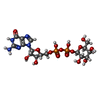
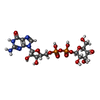


| #2: Chemical |  Mass: 605.341 Da / Num. of mol.: 2 / Source method: obtained synthetically / Formula: C16H25N5O16P2 Mass: 605.341 Da / Num. of mol.: 2 / Source method: obtained synthetically / Formula: C16H25N5O16P2#3: Chemical |  Mass: 603.325 Da / Num. of mol.: 2 / Source method: obtained synthetically / Formula: C16H23N5O16P2 Mass: 603.325 Da / Num. of mol.: 2 / Source method: obtained synthetically / Formula: C16H23N5O16P2#4: Chemical |  Mass: 663.425 Da / Num. of mol.: 2 / Source method: obtained synthetically / Formula: C21H27N7O14P2 / Comment: NAD*YM Mass: 663.425 Da / Num. of mol.: 2 / Source method: obtained synthetically / Formula: C21H27N7O14P2 / Comment: NAD*YM#5: Chemical | ChemComp-FMT /  Mass: 46.025 Da / Num. of mol.: 18 / Source method: obtained synthetically / Formula: CH2O2 Mass: 46.025 Da / Num. of mol.: 18 / Source method: obtained synthetically / Formula: CH2O2#6: Chemical | ChemComp-EPE / |  Mass: 238.305 Da / Num. of mol.: 1 / Source method: obtained synthetically / Formula: C8H18N2O4S / Comment: pH buffer*YM Mass: 238.305 Da / Num. of mol.: 1 / Source method: obtained synthetically / Formula: C8H18N2O4S / Comment: pH buffer*YM#7: Water | ChemComp-HOH / | Mass: 18.015 Da / Num. of mol.: 944 / Source method: isolated from a natural source / Formula: H2O |
|---|
-Details
| Compound details | ENGINEERED| Sequence details | THE FIRST TWO RESIDUES (GA) IN THE SPECIFIED SEQUENCE ARE A REMNANT FROM A CLEAVED HIS-TAG (AND ARE ...THE FIRST TWO RESIDUES (GA) IN THE SPECIFIED SEQUENCE ARE A REMNANT FROM A CLEAVED HIS-TAG (AND ARE NOT IN THE UNIPROT SEQUENCE), THE RESIDUE NUMBERING IN THE STRUCTURE USES THE THIRD RESIDUE (M) AS RESIDUE 1. | |
|---|
-Experimental details
-Experiment
| Experiment | Method: X-RAY DIFFRACTION / Number of used crystals: 1 |
|---|
- Sample preparation
| Crystal | Density Matthews: 1.95 Å3/Da / Density % sol: 37 % Description: INITIAL MODEL FOR MOLECULAR REPLACEMENT BY PHASER DETERMINED BY MAD OF SELENO-METHIONINE PROTEIN. DATA COLLECTED ON BM14, ANALYSED WITH SOLVE AND RESOLVE. |
|---|---|
| Crystal grow | Method: vapor diffusion, sitting drop / pH: 7.4 Details: PROTEIN CRYSTALISED IN 100MM HEPES PH 7.4, 2.16 M AMMONIUM SULPHATE, VAPOUR DIFFUSION, SITTING DROP. CRYOPROTECTED WITH 4M SODIUM FORMATE. |
-Data collection
| Diffraction | Mean temperature: 100 K |
|---|---|
| Diffraction source | Source: SYNCHROTRON / Site: ESRF  / Beamline: ID14-1 / Wavelength: 0.934 / Beamline: ID14-1 / Wavelength: 0.934 |
| Detector | Type: ADSC CCD / Detector: CCD / Date: Mar 13, 2004 |
| Radiation | Protocol: SINGLE WAVELENGTH / Monochromatic (M) / Laue (L): M / Scattering type: x-ray |
| Radiation wavelength | Wavelength: 0.934 Å / Relative weight: 1 |
| Reflection | Resolution: 1.5→23.3 Å / Num. obs: 105071 / % possible obs: 99.9 % / Redundancy: 3.7 % / Biso Wilson estimate: 13.37 Å2 / Rmerge(I) obs: 0.05 / Net I/σ(I): 9.6 |
| Reflection shell | Resolution: 1.5→1.54 Å / Redundancy: 3.6 % / Rmerge(I) obs: 0.21 / Mean I/σ(I) obs: 3.6 / % possible all: 99.9 |
- Processing
| Software |
| ||||||||||||||||||||||||||||||||||||||||||||||||||||||||||||||||||||||||||||||||||||||||||||||||||||||||||||||||||||||||||||||||||||||||||||||||||||||||||||||||||||||||||||||||||||||
|---|---|---|---|---|---|---|---|---|---|---|---|---|---|---|---|---|---|---|---|---|---|---|---|---|---|---|---|---|---|---|---|---|---|---|---|---|---|---|---|---|---|---|---|---|---|---|---|---|---|---|---|---|---|---|---|---|---|---|---|---|---|---|---|---|---|---|---|---|---|---|---|---|---|---|---|---|---|---|---|---|---|---|---|---|---|---|---|---|---|---|---|---|---|---|---|---|---|---|---|---|---|---|---|---|---|---|---|---|---|---|---|---|---|---|---|---|---|---|---|---|---|---|---|---|---|---|---|---|---|---|---|---|---|---|---|---|---|---|---|---|---|---|---|---|---|---|---|---|---|---|---|---|---|---|---|---|---|---|---|---|---|---|---|---|---|---|---|---|---|---|---|---|---|---|---|---|---|---|---|---|---|---|---|
| Refinement | Method to determine structure: MOLECULAR REPLACEMENT / Resolution: 1.5→64.55 Å / Cor.coef. Fo:Fc: 0.978 / Cor.coef. Fo:Fc free: 0.959 / SU B: 2.281 / SU ML: 0.04 / Cross valid method: THROUGHOUT / σ(F): 0.96 / ESU R: 0.082 / ESU R Free: 0.069 / Stereochemistry target values: RESTRAINED Details: HYDROGENS HAVE BEEN ADDED IN THE RIDING POSITIONS. EACH MONOMER OF GME K178R CONTAINS A MIXTURE OF COMPOUNDS IN THE ACTIVE SITE -NAD AND NADH IN THE NUCLEOTIDE BINDING SITE, GDP-BETA-L- ...Details: HYDROGENS HAVE BEEN ADDED IN THE RIDING POSITIONS. EACH MONOMER OF GME K178R CONTAINS A MIXTURE OF COMPOUNDS IN THE ACTIVE SITE -NAD AND NADH IN THE NUCLEOTIDE BINDING SITE, GDP-BETA-L-GULOSE AND GDP-BETA-L-4- KETO-GULOSE IN THE NUCLEOTIDE SUGAR BINDING SITE. DURING REFINEMENT THE CONSTANT PORTION OF EACH MOLECULE (ADP FOR NAD, GMP FOR THE GDP-SUGARS, AS THE SECOND PHOSPHATE GROUP MOVES WITH DIFFERENT LIGANDS) WAS FIXED WITH OCCUPANCY OF 1 AND LINKED TO TWO VARIABLE PORTIONS WITH PARTIAL OCCUPANCY.
| ||||||||||||||||||||||||||||||||||||||||||||||||||||||||||||||||||||||||||||||||||||||||||||||||||||||||||||||||||||||||||||||||||||||||||||||||||||||||||||||||||||||||||||||||||||||
| Solvent computation | Ion probe radii: 0.8 Å / Shrinkage radii: 0.8 Å / VDW probe radii: 1.4 Å / Solvent model: MASK BULK SOLVENT | ||||||||||||||||||||||||||||||||||||||||||||||||||||||||||||||||||||||||||||||||||||||||||||||||||||||||||||||||||||||||||||||||||||||||||||||||||||||||||||||||||||||||||||||||||||||
| Displacement parameters | Biso mean: 11.09 Å2
| ||||||||||||||||||||||||||||||||||||||||||||||||||||||||||||||||||||||||||||||||||||||||||||||||||||||||||||||||||||||||||||||||||||||||||||||||||||||||||||||||||||||||||||||||||||||
| Refinement step | Cycle: LAST / Resolution: 1.5→64.55 Å
| ||||||||||||||||||||||||||||||||||||||||||||||||||||||||||||||||||||||||||||||||||||||||||||||||||||||||||||||||||||||||||||||||||||||||||||||||||||||||||||||||||||||||||||||||||||||
| Refine LS restraints |
|