 Movie
Movie Controller

[English] 日本語
 Yorodumi
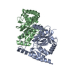
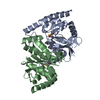
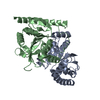
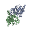
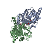
- PDB-7l9w: Wild-type Pseudomonas fluorescens isocyanide hydratase (WT-3) at ... -
+ Open data

- Basic information
| Entry | Database: PDB / ID: 7l9w | |||||||||
|---|---|---|---|---|---|---|---|---|---|---|
| Title | Wild-type Pseudomonas fluorescens isocyanide hydratase (WT-3) at 274K, Refmac5-refined | |||||||||
 Components Components | Isonitrile hydratase InhA | |||||||||
 Keywords Keywords | LYASE / DJ-1/PfpI superfamily | |||||||||
| Function / homology | : / DJ-1/PfpI / DJ-1/PfpI family / Class I glutamine amidotransferase-like / regulation of DNA-templated transcription / Isonitrile hydratase InhA Function and homology information Function and homology information | |||||||||
| Biological species |  Pseudomonas fluorescens (bacteria) Pseudomonas fluorescens (bacteria) | |||||||||
| Method |  X-RAY DIFFRACTION / SYNCHROTRON / MOLECULAR REPLACEMENT / Resolution: 1.199 Å X-RAY DIFFRACTION / SYNCHROTRON / MOLECULAR REPLACEMENT / Resolution: 1.199 Å | |||||||||
 Authors Authors | Su, Z. / Dasgupta, M. / Poitevin, F. / Mathews, I.I. / van den Bedem, H. / Wall, M.E. / Yoon, C.H. / Wilson, M.A. | |||||||||
| Funding support |  United States, 2items United States, 2items
| |||||||||
 Citation Citation | Journal: Struct Dyn. / Year: 2021 Title: Reproducibility of protein x-ray diffuse scattering and potential utility for modeling atomic displacement parameters. Authors: Su, Z. / Dasgupta, M. / Poitevin, F. / Mathews, I.I. / van den Bedem, H. / Wall, M.E. / Yoon, C.H. / Wilson, M.A. | |||||||||
| History |
|
- Structure visualization
| Structure viewer | Molecule: MolmilJmol/JSmol |
|---|
- Downloads & links
-Download
| PDBx/mmCIF format | 7l9w.cif.gz | 204.5 KB | Display | PDBx/mmCIF format |
|---|---|---|---|---|
| PDB format | pdb7l9w.ent.gz | 160.9 KB | Display | PDB format |
| PDBx/mmJSON format | 7l9w.json.gz | Tree view | PDBx/mmJSON format | |
| Others |  Other downloads Other downloads |
-Validation report
| Arichive directory | https://data.pdbj.org/pub/pdb/validation_reports/l9/7l9wftp://data.pdbj.org/pub/pdb/validation_reports/l9/7l9w | HTTPS FTP |
|---|
-Related structure data
| Related structure data |  7l9qC  7l9sC  7l9zC  7la0C 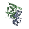 7la3C  7lavC  7laxC  7lb9C  7lbhC 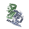 7lbiC 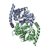 7lcxC 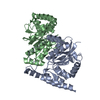 7ld6C 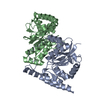 7ld7C  7ldbC 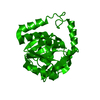 7ldiC  7ldmC 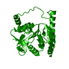 7ldoC 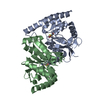 6ni6S C: citing same article ( S: Starting model for refinement |
|---|---|
| Similar structure data |


-Links
 PDBj
PDBj

- Assembly
| Deposited unit | 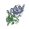
| ||||||||
|---|---|---|---|---|---|---|---|---|---|
| 1 |
| ||||||||
| Unit cell |
|
-Components
| #1: Protein | Mass: 24180.646 Da / Num. of mol.: 2 Source method: isolated from a genetically manipulated source Source: (gene. exp.) Pseudomonas fluorescens (strain ATCC BAA-477 / NRRL B-23932 / Pf-5) (bacteria)Strain: ATCC BAA-477 / NRRL B-23932 / Pf-5 / Gene: inhA, PFL_4109 / Plasmid: pET15b / Production host: #2: Water | ChemComp-HOH / |  Mass: 18.015 Da / Num. of mol.: 355 / Source method: isolated from a natural source / Formula: H2O Mass: 18.015 Da / Num. of mol.: 355 / Source method: isolated from a natural source / Formula: H2O |
|---|
-Experimental details
-Experiment
| Experiment | Method: X-RAY DIFFRACTION / Number of used crystals: 1 |
|---|
- Sample preparation
| Crystal | Density Matthews: 2.19 Å3/Da / Density % sol: 43.96 % |
|---|---|
| Crystal grow | Temperature: 298 K / Method: vapor diffusion, hanging drop / pH: 8.6 Details: 23% PEG 3350, 100MM TRIS-HCL PH 8.6, 200 MM MAGNESIUM CHLORIDE AND 2 MM DITHIOTHREITOL |
-Data collection
| Diffraction | Mean temperature: 274 K / Serial crystal experiment: N |
|---|---|
| Diffraction source | Source: SYNCHROTRON / Site: SSRL / Beamline: BL12-2 / Wavelength: 0.775 Å |
| Detector | Type: DECTRIS PILATUS 6M / Detector: PIXEL / Date: Nov 28, 2018 Details: Flat Si Rh coated M0, Kirkpatrick-Baez flat bent Si M1 & M2 |
| Radiation | Monochromator: Liquid nitrogen-cooled double crystal Si(111 / Protocol: SINGLE WAVELENGTH / Monochromatic (M) / Laue (L): M / Scattering type: x-ray |
| Radiation wavelength | Wavelength: 0.775 Å / Relative weight: 1 |
| Reflection | Resolution: 1.199→39.152 Å / Num. obs: 126107 / % possible obs: 96.5 % / Redundancy: 3.7 % / CC1/2: 0.997 / Rrim(I) all: 0.069 / Net I/σ(I): 7.1 |
| Reflection shell | Resolution: 1.199→1.22 Å / Redundancy: 3.5 % / Mean I/σ(I) obs: 1.1 / Num. unique obs: 5967 / CC1/2: 0.309 / Rrim(I) all: 1.737 / % possible all: 91.9 |
- Processing
| Software |
| ||||||||||||||||||||||||||||||||||||||||||||||||||||||||||||||||||||||||||||||||||||||||||||||||||||||||||||||||||||||||||||||||||||||||||||||||||||||||||||||||
|---|---|---|---|---|---|---|---|---|---|---|---|---|---|---|---|---|---|---|---|---|---|---|---|---|---|---|---|---|---|---|---|---|---|---|---|---|---|---|---|---|---|---|---|---|---|---|---|---|---|---|---|---|---|---|---|---|---|---|---|---|---|---|---|---|---|---|---|---|---|---|---|---|---|---|---|---|---|---|---|---|---|---|---|---|---|---|---|---|---|---|---|---|---|---|---|---|---|---|---|---|---|---|---|---|---|---|---|---|---|---|---|---|---|---|---|---|---|---|---|---|---|---|---|---|---|---|---|---|---|---|---|---|---|---|---|---|---|---|---|---|---|---|---|---|---|---|---|---|---|---|---|---|---|---|---|---|---|---|---|---|---|
| Refinement | Method to determine structure: MOLECULAR REPLACEMENT Starting model: 6NI6 Resolution: 1.199→39.152 Å / Cor.coef. Fo:Fc: 0.985 / Cor.coef. Fo:Fc free: 0.978 / SU B: 1.466 / SU ML: 0.027 / Cross valid method: FREE R-VALUE / ESU R: 0.035 / ESU R Free: 0.035 Details: Hydrogens have been added in their riding positions
| ||||||||||||||||||||||||||||||||||||||||||||||||||||||||||||||||||||||||||||||||||||||||||||||||||||||||||||||||||||||||||||||||||||||||||||||||||||||||||||||||
| Solvent computation | Ion probe radii: 0.8 Å / Shrinkage radii: 0.8 Å / VDW probe radii: 1.2 Å / Solvent model: MASK BULK SOLVENT | ||||||||||||||||||||||||||||||||||||||||||||||||||||||||||||||||||||||||||||||||||||||||||||||||||||||||||||||||||||||||||||||||||||||||||||||||||||||||||||||||
| Displacement parameters | Biso mean: 17.448 Å2
| ||||||||||||||||||||||||||||||||||||||||||||||||||||||||||||||||||||||||||||||||||||||||||||||||||||||||||||||||||||||||||||||||||||||||||||||||||||||||||||||||
| Refinement step | Cycle: LAST / Resolution: 1.199→39.152 Å
| ||||||||||||||||||||||||||||||||||||||||||||||||||||||||||||||||||||||||||||||||||||||||||||||||||||||||||||||||||||||||||||||||||||||||||||||||||||||||||||||||
| Refine LS restraints |
| ||||||||||||||||||||||||||||||||||||||||||||||||||||||||||||||||||||||||||||||||||||||||||||||||||||||||||||||||||||||||||||||||||||||||||||||||||||||||||||||||
| LS refinement shell |
|
