 Movie
Movie Controller


+ Open data

- Basic information
| Entry | Database: PDB / ID: 7l78 | ||||||
|---|---|---|---|---|---|---|---|
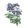
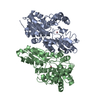
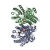
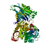
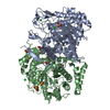
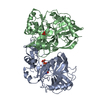
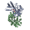
| Title | H235C variant of Yeast Ferrochelatase | ||||||
 Components Components | Ferrochelatase, mitochondrial | ||||||
 Keywords Keywords | LYASE / ferrochelatase / heme biosynthesis / yeast | ||||||
| Function / homology |  Function and homology information Function and homology informationHeme biosynthesis / protoporphyrin ferrochelatase / protoporphyrin ferrochelatase activity / Mitochondrial protein degradation / heme biosynthetic process / mitochondrial inner membrane / mitochondrion Similarity search - Function | ||||||
| Biological species |  | ||||||
| Method |  X-RAY DIFFRACTION / SYNCHROTRON / MOLECULAR REPLACEMENT / Resolution: 2.4 Å X-RAY DIFFRACTION / SYNCHROTRON / MOLECULAR REPLACEMENT / Resolution: 2.4 Å | ||||||
 Authors Authors | Lanzilotta, W.N. / Medlock, A.E. | ||||||
| Funding support | 1items
| ||||||
 Citation Citation | Journal: Redox Biol / Year: 2021 Title: Mitochondrial contact site and cristae organizing system (MICOS) machinery supports heme biosynthesis by enabling optimal performance of ferrochelatase. Authors: Dietz, J.V. / Willoughby, M.M. / Piel III, R.B. / Ross, T.A. / Bohovych, I. / Addis, H.G. / Fox, J.L. / Lanzilotta, W.N. / Dailey, H.A. / Wohlschlegel, J.A. / Reddi, A.R. / Medlock, A.E. / Khalimonchuk, O. | ||||||
| History |
|
- Structure visualization
| Structure viewer | Molecule: MolmilJmol/JSmol |
|---|
- Downloads & links
-Download
| PDBx/mmCIF format | 7l78.cif.gz | 155.1 KB | Display | PDBx/mmCIF format |
|---|---|---|---|---|
| PDB format | pdb7l78.ent.gz | 121 KB | Display | PDB format |
| PDBx/mmJSON format | 7l78.json.gz | Tree view | PDBx/mmJSON format | |
| Others |  Other downloads Other downloads |
-Validation report
| Arichive directory | https://data.pdbj.org/pub/pdb/validation_reports/l7/7l78ftp://data.pdbj.org/pub/pdb/validation_reports/l7/7l78 | HTTPS FTP |
|---|
-Related structure data
| Related structure data |  1lbqS S: Starting model for refinement |
|---|---|
| Similar structure data |


-Links
 PDBj
PDBj
- Assembly
| Deposited unit | 
| ||||||||
|---|---|---|---|---|---|---|---|---|---|
| 1 |
| ||||||||
| Unit cell |
|
-Components
| #1: Protein | Mass: 40334.988 Da / Num. of mol.: 2 Source method: isolated from a genetically manipulated source Source: (gene. exp.) Gene: HEM15, YOR176W, HemH / Production host:  #2: Chemical | 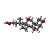  Mass: 408.571 Da / Num. of mol.: 2 / Source method: obtained synthetically / Formula: C24H40O5 Mass: 408.571 Da / Num. of mol.: 2 / Source method: obtained synthetically / Formula: C24H40O5#3: Water | ChemComp-HOH / |  Mass: 18.015 Da / Num. of mol.: 63 / Source method: isolated from a natural source / Formula: H2O Mass: 18.015 Da / Num. of mol.: 63 / Source method: isolated from a natural source / Formula: H2OHas ligand of interest | N | |
|---|
-Experimental details
-Experiment
| Experiment | Method: X-RAY DIFFRACTION / Number of used crystals: 1 |
|---|
- Sample preparation
| Crystal | Density Matthews: 2.3 Å3/Da / Density % sol: 46.58 % |
|---|---|
| Crystal grow | Temperature: 291 K / Method: vapor diffusion, sitting drop / Details: PEG 2000, 2-propanol, Tris-HCl pH 7.5 |
-Data collection
| Diffraction | Mean temperature: 100 K / Serial crystal experiment: N |
|---|---|
| Diffraction source | Source: SYNCHROTRON / Site: APS  / Beamline: 22-ID / Wavelength: 0.98 Å / Beamline: 22-ID / Wavelength: 0.98 Å |
| Detector | Type: DECTRIS PILATUS 300K / Detector: PIXEL / Date: Mar 20, 2013 |
| Radiation | Monochromator: 0.98 / Protocol: SINGLE WAVELENGTH / Monochromatic (M) / Laue (L): M / Scattering type: x-ray |
| Radiation wavelength | Wavelength: 0.98 Å / Relative weight: 1 |
| Reflection | Resolution: 2.4→50 Å / Num. obs: 32665 / % possible obs: 99.2 % / Redundancy: 3.2 % / CC1/2: 0.995 / Net I/σ(I): 17.7 |
| Reflection shell | Resolution: 2.4→2.52 Å / Num. unique obs: 3102 / CC1/2: 0.795 |
- Processing
| Software |
| ||||||||||||||||||||||||||||||||||||||||||||||||||||||||||||||||||||||||
|---|---|---|---|---|---|---|---|---|---|---|---|---|---|---|---|---|---|---|---|---|---|---|---|---|---|---|---|---|---|---|---|---|---|---|---|---|---|---|---|---|---|---|---|---|---|---|---|---|---|---|---|---|---|---|---|---|---|---|---|---|---|---|---|---|---|---|---|---|---|---|---|---|---|
| Refinement | Method to determine structure: MOLECULAR REPLACEMENT Starting model: 1LBQ Resolution: 2.4→41.926 Å / SU ML: 0.36 / Cross valid method: THROUGHOUT / σ(F): 1.35 / Phase error: 31.36 / Stereochemistry target values: ML
| ||||||||||||||||||||||||||||||||||||||||||||||||||||||||||||||||||||||||
| Solvent computation | Shrinkage radii: 0.9 Å / VDW probe radii: 1.11 Å / Solvent model: FLAT BULK SOLVENT MODEL | ||||||||||||||||||||||||||||||||||||||||||||||||||||||||||||||||||||||||
| Displacement parameters | Biso max: 110.21 Å2 / Biso mean: 48.2448 Å2 / Biso min: 19.03 Å2 | ||||||||||||||||||||||||||||||||||||||||||||||||||||||||||||||||||||||||
| Refinement step | Cycle: final / Resolution: 2.4→41.926 Å
| ||||||||||||||||||||||||||||||||||||||||||||||||||||||||||||||||||||||||
| LS refinement shell | Refine-ID: X-RAY DIFFRACTION / Rfactor Rfree error: 0
|
