 Movie
Movie Controller

[English] 日本語
 Yorodumi
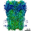
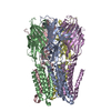
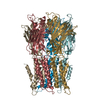
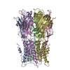
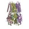
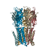
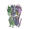
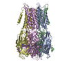
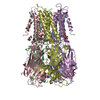
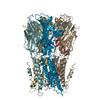
- PDB-7l6q: Unliganded ELIC in styrene-maleic-acid nanodiscs at 2.5-Angstrom ... -
+ Open data

- Basic information
| Entry | Database: PDB / ID: 7l6q | ||||||
|---|---|---|---|---|---|---|---|
| Title | Unliganded ELIC in styrene-maleic-acid nanodiscs at 2.5-Angstrom resolution | ||||||
 Components Components | Gamma-aminobutyric-acid receptor subunit beta-1 | ||||||
 Keywords Keywords | MEMBRANE PROTEIN / Pentameric Ligand-gated Ion Channels / Nanodisc / Cys-loop receptor / Styrene-maleic acid / Protein-lipid interface | ||||||
| Function / homology |  Function and homology information Function and homology informationextracellular ligand-gated monoatomic ion channel activity / transmembrane signaling receptor activity / identical protein binding / plasma membrane Similarity search - Function | ||||||
| Biological species |  Dickeya dadantii (bacteria) Dickeya dadantii (bacteria) | ||||||
| Method | ELECTRON MICROSCOPY / single particle reconstruction / cryo EM / Resolution: 2.5 Å | ||||||
 Authors Authors | Grosman, C. / Kumar, P. | ||||||
| Funding support |  United States, 1items United States, 1items
| ||||||
 Citation Citation | Journal: Proc Natl Acad Sci U S A / Year: 2021 Title: Structure and function at the lipid-protein interface of a pentameric ligand-gated ion channel. Authors: Pramod Kumar / Gisela D Cymes / Claudio Grosman / Abstract: Although it has long been proposed that membrane proteins may contain tightly bound lipids, their identity, the structure of their binding sites, and their functional and structural relevance have ...Although it has long been proposed that membrane proteins may contain tightly bound lipids, their identity, the structure of their binding sites, and their functional and structural relevance have remained elusive. To some extent, this is because tightly bound lipids are often located at the periphery of proteins, where the quality of density maps is usually poorer, and because they may be outcompeted by detergent molecules used during standard purification procedures. As a step toward characterizing natively bound lipids in the superfamily of pentameric ligand-gated ion channels (pLGICs), we applied single-particle cryogenic electron microscopy to fragments of native membrane obtained in the complete absence of detergent-solubilization steps. Because of the heterogeneous lipid composition of membranes in the secretory pathway of eukaryotic cells, we chose to study a bacterial pLGIC (ELIC) expressed in 's inner membrane. We obtained a three-dimensional reconstruction of unliganded ELIC (2.5-Å resolution) that shows clear evidence for two types of tightly bound lipid at the protein-bulk-membrane interface. One of them was consistent with a "regular" diacylated phospholipid, in the cytoplasmic leaflet, whereas the other one was consistent with the tetra-acylated structure of cardiolipin, in the periplasmic leaflet. Upon reconstitution in polar-lipid bilayers, ELIC retained the functional properties characteristic of members of this superfamily, and thus, the fitted atomic model is expected to represent the (long-debated) unliganded-closed, "resting" conformation of this ion channel. Notably, the addition of cardiolipin to phosphatidylcholine membranes restored the ion-channel activity that is largely lost in phosphatidylcholine-only bilayers. | ||||||
| History |
|
- Structure visualization
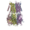
| Movie |
Movie viewer |
|---|---|
| Structure viewer | Molecule: MolmilJmol/JSmol |
- Downloads & links
-Download
| PDBx/mmCIF format | 7l6q.cif.gz | 293.1 KB | Display | PDBx/mmCIF format |
|---|---|---|---|---|
| PDB format | pdb7l6q.ent.gz | 241.7 KB | Display | PDB format |
| PDBx/mmJSON format | 7l6q.json.gz | Tree view | PDBx/mmJSON format | |
| Others |  Other downloads Other downloads |
-Validation report
| Arichive directory | https://data.pdbj.org/pub/pdb/validation_reports/l6/7l6qftp://data.pdbj.org/pub/pdb/validation_reports/l6/7l6q | HTTPS FTP |
|---|
-Related structure data
| Related structure data | 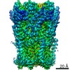 23207MC 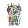 7l6uC M: map data used to model this data C: citing same article ( |
|---|---|
| Similar structure data |
-Links
 PDBj
PDBj


- Assembly
| Deposited unit | 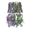
| |||||||||||||||||||||||||||||||||||||||||||||||||||||||||||||||||||||||||||||||||||||||||||||||
|---|---|---|---|---|---|---|---|---|---|---|---|---|---|---|---|---|---|---|---|---|---|---|---|---|---|---|---|---|---|---|---|---|---|---|---|---|---|---|---|---|---|---|---|---|---|---|---|---|---|---|---|---|---|---|---|---|---|---|---|---|---|---|---|---|---|---|---|---|---|---|---|---|---|---|---|---|---|---|---|---|---|---|---|---|---|---|---|---|---|---|---|---|---|---|---|---|
| 1 |
| |||||||||||||||||||||||||||||||||||||||||||||||||||||||||||||||||||||||||||||||||||||||||||||||
| Noncrystallographic symmetry (NCS) | NCS domain:
NCS domain segments:
|
-Components
| #1: Protein | Mass: 36879.000 Da / Num. of mol.: 5 Source method: isolated from a genetically manipulated source Source: (gene. exp.) Dickeya dadantii (strain 3937) (bacteria)Strain: 3937 / Gene: Dda3937_00520 / Production host: #2: Chemical | ChemComp-PGW / ( 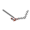  Mass: 749.007 Da / Num. of mol.: 5 / Source method: obtained synthetically / Formula: C40H77O10P / Comment: phospholipid*YM Mass: 749.007 Da / Num. of mol.: 5 / Source method: obtained synthetically / Formula: C40H77O10P / Comment: phospholipid*YM#3: Chemical | ChemComp-CDL /   Mass: 1464.043 Da / Num. of mol.: 5 / Source method: obtained synthetically / Formula: C81H156O17P2 / Feature type: SUBJECT OF INVESTIGATION / Comment: phospholipid*YM Mass: 1464.043 Da / Num. of mol.: 5 / Source method: obtained synthetically / Formula: C81H156O17P2 / Feature type: SUBJECT OF INVESTIGATION / Comment: phospholipid*YMHas ligand of interest | Y | |
|---|
-Experimental details
-Experiment
| Experiment | Method: ELECTRON MICROSCOPY |
|---|---|
| EM experiment | Aggregation state: PARTICLE / 3D reconstruction method: single particle reconstruction |
- Sample preparation
| Component | Name: Unliganded Erwinia chrysanthemi ligand gated ion channel in styrene-maleic-acid nanodiscs Type: COMPLEX / Entity ID: #1 / Source: RECOMBINANT | |||||||||||||||
|---|---|---|---|---|---|---|---|---|---|---|---|---|---|---|---|---|
| Molecular weight | Value: 184.2 kDa/nm / Experimental value: NO | |||||||||||||||
| Source (natural) | Organism: Dickeya dadantii (bacteria) | |||||||||||||||
| Source (recombinant) | Organism: Strain: BL21 | |||||||||||||||
| Buffer solution | pH: 8 | |||||||||||||||
| Buffer component |
| |||||||||||||||
| Specimen | Embedding applied: NO / Shadowing applied: NO / Staining applied: NO / Vitrification applied: YES | |||||||||||||||
| Specimen support | Grid material: GOLD | |||||||||||||||
| Vitrification | Instrument: LEICA EM GP / Cryogen name: ETHANE |
- Electron microscopy imaging
| Experimental equipment |  Model: Titan Krios / Image courtesy: FEI Company |
|---|---|
| Microscopy | Model: FEI TITAN KRIOS |
| Electron gun | Electron source:  FIELD EMISSION GUN / Accelerating voltage: 300 kV / Illumination mode: SPOT SCAN FIELD EMISSION GUN / Accelerating voltage: 300 kV / Illumination mode: SPOT SCAN |
| Electron lens | Mode: BRIGHT FIELD |
| Specimen holder | Specimen holder model: FEI TITAN KRIOS AUTOGRID HOLDER |
| Image recording | Electron dose: 56.23 e/Å2 / Film or detector model: GATAN K2 SUMMIT (4k x 4k) |
- Processing
| Software | Name: PHENIX / Version: 1.18.2_3874: / Classification: refinement | ||||||||||||||||||||||||||||||
|---|---|---|---|---|---|---|---|---|---|---|---|---|---|---|---|---|---|---|---|---|---|---|---|---|---|---|---|---|---|---|---|
| CTF correction | Type: NONE | ||||||||||||||||||||||||||||||
| Symmetry | Point symmetry: C5 (5 fold cyclic) | ||||||||||||||||||||||||||||||
| 3D reconstruction | Resolution: 2.5 Å / Resolution method: FSC 0.143 CUT-OFF / Num. of particles: 1239532 / Symmetry type: POINT | ||||||||||||||||||||||||||||||
| Refinement | Cross valid method: NONE Stereochemistry target values: GeoStd + Monomer Library + CDL v1.2 | ||||||||||||||||||||||||||||||
| Displacement parameters | Biso mean: 61.5 Å2 | ||||||||||||||||||||||||||||||
| Refine LS restraints |
| ||||||||||||||||||||||||||||||
| Refine LS restraints NCS |
|
