 Movie
Movie Controller

[English] 日本語
 Yorodumi
- PDB-7l5r: Crystal Structure of the Oxacillin-hydrolyzing Class D Extended-s... -
+ Open data

- Basic information
| Entry | Database: PDB / ID: 7l5r | ||||||
|---|---|---|---|---|---|---|---|
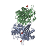
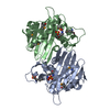
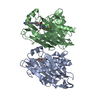
| Title | Crystal Structure of the Oxacillin-hydrolyzing Class D Extended-spectrum Beta-lactamase OXA-14 from Pseudomonas aeruginosa | ||||||
 Components Components | (Beta-lactamase) x 2 | ||||||
 Keywords Keywords | HYDROLASE / Structural Genomics / Center for Structural Genomics of Infectious Diseases / CSGID / class D beta-lactamase / OXA-14 | ||||||
| Function / homology |  Function and homology information Function and homology informationpenicillin binding / antibiotic catabolic process / cell wall organization / beta-lactamase activity / beta-lactamase / response to antibiotic / plasma membrane Similarity search - Function | ||||||
| Biological species |   Pseudomonas aeruginosa (bacteria) Pseudomonas aeruginosa (bacteria) | ||||||
| Method |  X-RAY DIFFRACTION / SYNCHROTRON / MOLECULAR REPLACEMENT / Resolution: 1.65 Å X-RAY DIFFRACTION / SYNCHROTRON / MOLECULAR REPLACEMENT / Resolution: 1.65 Å | ||||||
 Authors Authors | Minasov, G. / Shuvalova, L. / Rosas-Lemus, M. / Brunzelle, J.S. / Satchell, K.J.F. / Center for Structural Genomics of Infectious Diseases (CSGID) | ||||||
 Citation Citation | Journal: Antimicrob.Agents Chemother. / Year: 2022 Title: Functional and Structural Characterization of OXA-935, a Novel OXA-10-Family beta-Lactamase from Pseudomonas aeruginosa. Authors: Pincus, N.B. / Rosas-Lemus, M. / Gatesy, S.W.M. / Bertucci, H.K. / Brunzelle, J.S. / Minasov, G. / Shuvalova, L.A. / Lebrun-Corbin, M. / Satchell, K.J.F. / Ozer, E.A. / Hauser, A.R. / Bachta, K.E.R. | ||||||
| History |
|
- Structure visualization
| Structure viewer | Molecule: MolmilJmol/JSmol |
|---|
- Downloads & links
-Download
| PDBx/mmCIF format | 7l5r.cif.gz | 223 KB | Display | PDBx/mmCIF format |
|---|---|---|---|---|
| PDB format | pdb7l5r.ent.gz | 176.6 KB | Display | PDB format |
| PDBx/mmJSON format | 7l5r.json.gz | Tree view | PDBx/mmJSON format | |
| Others |  Other downloads Other downloads |
-Validation report
| Summary document | 7l5r_validation.pdf.gz | 464.8 KB | Display | wwPDB validaton report |
|---|---|---|---|---|
| Full document | 7l5r_full_validation.pdf.gz | 468 KB | Display | |
| Data in XML | 7l5r_validation.xml.gz | 25.2 KB | Display | |
| Data in CIF | 7l5r_validation.cif.gz | 37.6 KB | Display | |
| Arichive directory | https://data.pdbj.org/pub/pdb/validation_reports/l5/7l5rftp://data.pdbj.org/pub/pdb/validation_reports/l5/7l5r | HTTPS FTP |
-Related structure data
| Related structure data | 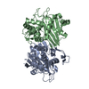 7l5vC  7n1mC 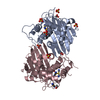 1e3uS S: Starting model for refinement C: citing same article ( |
|---|---|
| Similar structure data | |
| Other databases |


-Links
 PDBj
PDBj- Assembly
| Deposited unit | 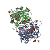
| ||||||||
|---|---|---|---|---|---|---|---|---|---|
| 1 |
| ||||||||
| Unit cell |
|
-Components
| #1: Protein | Mass: 27826.512 Da / Num. of mol.: 1 Source method: isolated from a genetically manipulated source Details: The side chain of Lys-70 is N-carboxylated / Source: (gene. exp.) Pseudomonas aeruginosa (bacteria) / Gene: OXA-14, blaOXA / Plasmid: pMCSG53 / Production host: | ||||||
|---|---|---|---|---|---|---|---|
| #2: Protein | Mass: 27783.510 Da / Num. of mol.: 1 Source method: isolated from a genetically manipulated source Source: (gene. exp.) Pseudomonas aeruginosa (bacteria) / Gene: OXA-14, blaOXA / Plasmid: pMCSG53 / Production host: | ||||||
| #3: Chemical |   Mass: 92.094 Da / Num. of mol.: 3 / Source method: obtained synthetically / Formula: C3H8O3 Mass: 92.094 Da / Num. of mol.: 3 / Source method: obtained synthetically / Formula: C3H8O3#4: Chemical | ChemComp-SO4 /   Mass: 96.063 Da / Num. of mol.: 22 / Source method: obtained synthetically / Formula: SO4 Mass: 96.063 Da / Num. of mol.: 22 / Source method: obtained synthetically / Formula: SO4#5: Water | ChemComp-HOH / |  Mass: 18.015 Da / Num. of mol.: 484 / Source method: isolated from a natural source / Formula: H2O Mass: 18.015 Da / Num. of mol.: 484 / Source method: isolated from a natural source / Formula: H2OHas ligand of interest | Y | |
-Experimental details
-Experiment
| Experiment | Method: X-RAY DIFFRACTION / Number of used crystals: 1 |
|---|
- Sample preparation
| Crystal | Density Matthews: 2.65 Å3/Da / Density % sol: 53.6 % |
|---|---|
| Crystal grow | Temperature: 292 K / Method: vapor diffusion, sitting drop / pH: 9 Details: Protein: 8.0 mg/ml, 0.01M Tris pH 8.3; Screen, AmSO4 (F6): 0.1M Bicine pH 9.0, 2.4M Ammonium sulfate; Cryo: 2M Lithium sulfate |
-Data collection
| Diffraction | Mean temperature: 100 K / Serial crystal experiment: N |
|---|---|
| Diffraction source | Source: SYNCHROTRON / Site: APS  / Beamline: 21-ID-F / Wavelength: 0.97872 Å / Beamline: 21-ID-F / Wavelength: 0.97872 Å |
| Detector | Type: MARMOSAIC 300 mm CCD / Detector: CCD / Date: May 13, 2020 / Details: Be |
| Radiation | Monochromator: Diamond / Protocol: SINGLE WAVELENGTH / Monochromatic (M) / Laue (L): M / Scattering type: x-ray |
| Radiation wavelength | Wavelength: 0.97872 Å / Relative weight: 1 |
| Reflection | Resolution: 1.65→30 Å / Num. obs: 71761 / % possible obs: 100 % / Observed criterion σ(I): -3 / Redundancy: 6 % / Biso Wilson estimate: 21.1 Å2 / CC1/2: 0.999 / CC star: 1 / Rmerge(I) obs: 0.06 / Rpim(I) all: 0.027 / Rrim(I) all: 0.066 / Rsym value: 0.06 / Χ2: 1.007 / Net I/σ(I): 27 |
| Reflection shell | Resolution: 1.65→1.68 Å / Redundancy: 6.1 % / Rmerge(I) obs: 0.841 / Mean I/σ(I) obs: 2.4 / Num. unique obs: 3530 / CC1/2: 0.8 / CC star: 0.943 / Rpim(I) all: 0.365 / Rrim(I) all: 0.919 / Rsym value: 0.841 / Χ2: 1.001 / % possible all: 100 |
- Processing
| Software |
| |||||||||||||||||||||||||||||||||||||||||||||||||||||||||||||||||||||||||||||||||||||||||||||||||||||||||||||||||||||||||||||||||||||||||||||||||||||||||||||||||||||||||||||||||||||||||||||||||||||||||||||||||||||||||||||||||||||||||||||||||||||||||||||||||||||||||||||||||||||||||||||||||||||||||||||||||||||||||||||||||||||
|---|---|---|---|---|---|---|---|---|---|---|---|---|---|---|---|---|---|---|---|---|---|---|---|---|---|---|---|---|---|---|---|---|---|---|---|---|---|---|---|---|---|---|---|---|---|---|---|---|---|---|---|---|---|---|---|---|---|---|---|---|---|---|---|---|---|---|---|---|---|---|---|---|---|---|---|---|---|---|---|---|---|---|---|---|---|---|---|---|---|---|---|---|---|---|---|---|---|---|---|---|---|---|---|---|---|---|---|---|---|---|---|---|---|---|---|---|---|---|---|---|---|---|---|---|---|---|---|---|---|---|---|---|---|---|---|---|---|---|---|---|---|---|---|---|---|---|---|---|---|---|---|---|---|---|---|---|---|---|---|---|---|---|---|---|---|---|---|---|---|---|---|---|---|---|---|---|---|---|---|---|---|---|---|---|---|---|---|---|---|---|---|---|---|---|---|---|---|---|---|---|---|---|---|---|---|---|---|---|---|---|---|---|---|---|---|---|---|---|---|---|---|---|---|---|---|---|---|---|---|---|---|---|---|---|---|---|---|---|---|---|---|---|---|---|---|---|---|---|---|---|---|---|---|---|---|---|---|---|---|---|---|---|---|---|---|---|---|---|---|---|---|---|---|---|---|---|---|---|---|---|---|---|---|---|---|---|---|---|---|---|---|---|---|---|---|---|---|---|---|---|---|---|---|---|---|---|---|---|---|---|---|---|---|---|---|---|---|---|---|---|---|---|---|---|---|---|
| Refinement | Method to determine structure: MOLECULAR REPLACEMENT Starting model: 1E3U Resolution: 1.65→29.75 Å / Cor.coef. Fo:Fc: 0.968 / Cor.coef. Fo:Fc free: 0.959 / SU B: 3.517 / SU ML: 0.058 / Cross valid method: THROUGHOUT / σ(F): 0 / ESU R: 0.083 / ESU R Free: 0.082 / Stereochemistry target values: MAXIMUM LIKELIHOOD Details: HYDROGENS HAVE BEEN ADDED IN THE RIDING POSITIONS U VALUES : WITH TLS ADDED
| |||||||||||||||||||||||||||||||||||||||||||||||||||||||||||||||||||||||||||||||||||||||||||||||||||||||||||||||||||||||||||||||||||||||||||||||||||||||||||||||||||||||||||||||||||||||||||||||||||||||||||||||||||||||||||||||||||||||||||||||||||||||||||||||||||||||||||||||||||||||||||||||||||||||||||||||||||||||||||||||||||||
| Solvent computation | Ion probe radii: 0.8 Å / Shrinkage radii: 0.8 Å / VDW probe radii: 1.2 Å / Solvent model: MASK | |||||||||||||||||||||||||||||||||||||||||||||||||||||||||||||||||||||||||||||||||||||||||||||||||||||||||||||||||||||||||||||||||||||||||||||||||||||||||||||||||||||||||||||||||||||||||||||||||||||||||||||||||||||||||||||||||||||||||||||||||||||||||||||||||||||||||||||||||||||||||||||||||||||||||||||||||||||||||||||||||||||
| Displacement parameters | Biso max: 105.72 Å2 / Biso mean: 25.851 Å2 / Biso min: 12.47 Å2
| |||||||||||||||||||||||||||||||||||||||||||||||||||||||||||||||||||||||||||||||||||||||||||||||||||||||||||||||||||||||||||||||||||||||||||||||||||||||||||||||||||||||||||||||||||||||||||||||||||||||||||||||||||||||||||||||||||||||||||||||||||||||||||||||||||||||||||||||||||||||||||||||||||||||||||||||||||||||||||||||||||||
| Refinement step | Cycle: final / Resolution: 1.65→29.75 Å
| |||||||||||||||||||||||||||||||||||||||||||||||||||||||||||||||||||||||||||||||||||||||||||||||||||||||||||||||||||||||||||||||||||||||||||||||||||||||||||||||||||||||||||||||||||||||||||||||||||||||||||||||||||||||||||||||||||||||||||||||||||||||||||||||||||||||||||||||||||||||||||||||||||||||||||||||||||||||||||||||||||||
| Refine LS restraints |
| |||||||||||||||||||||||||||||||||||||||||||||||||||||||||||||||||||||||||||||||||||||||||||||||||||||||||||||||||||||||||||||||||||||||||||||||||||||||||||||||||||||||||||||||||||||||||||||||||||||||||||||||||||||||||||||||||||||||||||||||||||||||||||||||||||||||||||||||||||||||||||||||||||||||||||||||||||||||||||||||||||||
| LS refinement shell | Resolution: 1.65→1.693 Å / Rfactor Rfree error: 0 / Total num. of bins used: 20
| |||||||||||||||||||||||||||||||||||||||||||||||||||||||||||||||||||||||||||||||||||||||||||||||||||||||||||||||||||||||||||||||||||||||||||||||||||||||||||||||||||||||||||||||||||||||||||||||||||||||||||||||||||||||||||||||||||||||||||||||||||||||||||||||||||||||||||||||||||||||||||||||||||||||||||||||||||||||||||||||||||||
| Refinement TLS params. | Method: refined / Refine-ID: X-RAY DIFFRACTION
| |||||||||||||||||||||||||||||||||||||||||||||||||||||||||||||||||||||||||||||||||||||||||||||||||||||||||||||||||||||||||||||||||||||||||||||||||||||||||||||||||||||||||||||||||||||||||||||||||||||||||||||||||||||||||||||||||||||||||||||||||||||||||||||||||||||||||||||||||||||||||||||||||||||||||||||||||||||||||||||||||||||
| Refinement TLS group |
|
