 Movie
Movie Controller


+ Open data

- Basic information
| Entry | Database: PDB / ID: 7l5k | ||||||
|---|---|---|---|---|---|---|---|
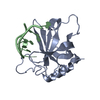
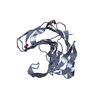
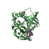
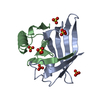
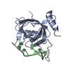
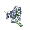
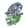
| Title | Crystal structure of the DiB-RM protein | ||||||
 Components Components | Lipocalin family protein | ||||||
 Keywords Keywords | FLUORESCENT PROTEIN / lipocalin / beta barrel / fluorogen activating protein / designed protein | ||||||
| Function / homology | ISOPROPYL ALCOHOL / :  Function and homology information Function and homology information | ||||||
| Biological species |  | ||||||
| Method |  X-RAY DIFFRACTION / SYNCHROTRON / MOLECULAR REPLACEMENT / molecular replacement / Resolution: 1.86 Å X-RAY DIFFRACTION / SYNCHROTRON / MOLECULAR REPLACEMENT / molecular replacement / Resolution: 1.86 Å | ||||||
 Authors Authors | Bozhanova, N.G. / Harp, J.M. / Meiler, J. | ||||||
| Funding support |  United States, 1items United States, 1items
| ||||||
 Citation Citation | Journal: Plos Comput.Biol. / Year: 2021 Title: Computational redesign of a fluorogen activating protein with Rosetta. Authors: Bozhanova, N.G. / Harp, J.M. / Bender, B.J. / Gavrikov, A.S. / Gorbachev, D.A. / Baranov, M.S. / Mercado, C.B. / Zhang, X. / Lukyanov, K.A. / Mishin, A.S. / Meiler, J. | ||||||
| History |
|
- Structure visualization
| Structure viewer | Molecule: MolmilJmol/JSmol |
|---|
- Downloads & links
-Download
| PDBx/mmCIF format | 7l5k.cif.gz | 88.3 KB | Display | PDBx/mmCIF format |
|---|---|---|---|---|
| PDB format | pdb7l5k.ent.gz | 63.3 KB | Display | PDB format |
| PDBx/mmJSON format | 7l5k.json.gz | Tree view | PDBx/mmJSON format | |
| Others |  Other downloads Other downloads |
-Validation report
| Arichive directory | https://data.pdbj.org/pub/pdb/validation_reports/l5/7l5kftp://data.pdbj.org/pub/pdb/validation_reports/l5/7l5k | HTTPS FTP |
|---|
-Related structure data
| Related structure data |  7l5lC 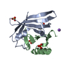 7l5mC 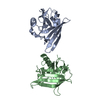 1qwdS S: Starting model for refinement C: citing same article ( |
|---|---|
| Similar structure data |


-Links
 PDBj
PDBj

- Assembly
| Deposited unit | 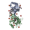
| ||||||||
|---|---|---|---|---|---|---|---|---|---|
| 1 | 
| ||||||||
| 2 | 
| ||||||||
| Unit cell |
|
-Components
| #1: Protein | Mass: 20184.699 Da / Num. of mol.: 2 / Fragment: UNP residues 20-177 / Mutation: P22S, A36C, F53A, N76F, S89Y, E90V, L141N Source method: isolated from a genetically manipulated source Source: (gene. exp.) #2: Chemical | ChemComp-IPA /   Mass: 60.095 Da / Num. of mol.: 4 / Source method: obtained synthetically / Formula: C3H8O Mass: 60.095 Da / Num. of mol.: 4 / Source method: obtained synthetically / Formula: C3H8O#3: Sugar | ChemComp-LMT /   Type: D-saccharide / Mass: 510.615 Da / Num. of mol.: 4 / Source method: obtained synthetically / Formula: C24H46O11 / Comment: detergent*YM Type: D-saccharide / Mass: 510.615 Da / Num. of mol.: 4 / Source method: obtained synthetically / Formula: C24H46O11 / Comment: detergent*YM#4: Chemical | ChemComp-SO4 /   Mass: 96.063 Da / Num. of mol.: 12 / Source method: obtained synthetically / Formula: SO4 Mass: 96.063 Da / Num. of mol.: 12 / Source method: obtained synthetically / Formula: SO4#5: Water | ChemComp-HOH / |  Mass: 18.015 Da / Num. of mol.: 169 / Source method: isolated from a natural source / Formula: H2O Mass: 18.015 Da / Num. of mol.: 169 / Source method: isolated from a natural source / Formula: H2OHas ligand of interest | N | |
|---|
-Experimental details
-Experiment
| Experiment | Method: X-RAY DIFFRACTION / Number of used crystals: 1 |
|---|
- Sample preparation
| Crystal | Density Matthews: 2.75 Å3/Da / Density % sol: 55.26 % |
|---|---|
| Crystal grow | Temperature: 294 K / Method: vapor diffusion, hanging drop Details: 2 M ammonium sulfate, 2.5% 2-propanol, 5% w/v n-Dodecyl-b-D-maltoside |
-Data collection
| Diffraction | Mean temperature: 100 K / Serial crystal experiment: N | ||||||||||||||||||||||||
|---|---|---|---|---|---|---|---|---|---|---|---|---|---|---|---|---|---|---|---|---|---|---|---|---|---|
| Diffraction source | Source: SYNCHROTRON / Site: APS / Beamline: 21-ID-F / Wavelength: 0.97872 Å | ||||||||||||||||||||||||
| Detector | Type: MARMOSAIC 225 mm CCD / Detector: CCD / Date: Aug 17, 2017 | ||||||||||||||||||||||||
| Radiation | Monochromator: diamond(111) / Protocol: SINGLE WAVELENGTH / Monochromatic (M) / Laue (L): M / Scattering type: x-ray | ||||||||||||||||||||||||
| Radiation wavelength | Wavelength: 0.97872 Å / Relative weight: 1 | ||||||||||||||||||||||||
| Reflection | Resolution: 1.86→33.32 Å / Num. obs: 36055 / % possible obs: 99.8 % / Redundancy: 4.1 % / Biso Wilson estimate: 35.517 Å2 / Rpim(I) all: 0.036 / Rrim(I) all: 0.077 / Net I/σ(I): 9.1 / Num. measured all: 148895 | ||||||||||||||||||||||||
| Reflection shell | Diffraction-ID: 1
|
-Phasing
| Phasing | Method: molecular replacement | ||||||
|---|---|---|---|---|---|---|---|
| Phasing MR | R rigid body: 0.354
|
- Processing
| Software |
| |||||||||||||||||||||||||||||||||||||||||||||||||||||||||||||||||||||||||||
|---|---|---|---|---|---|---|---|---|---|---|---|---|---|---|---|---|---|---|---|---|---|---|---|---|---|---|---|---|---|---|---|---|---|---|---|---|---|---|---|---|---|---|---|---|---|---|---|---|---|---|---|---|---|---|---|---|---|---|---|---|---|---|---|---|---|---|---|---|---|---|---|---|---|---|---|---|
| Refinement | Method to determine structure: MOLECULAR REPLACEMENT Starting model: PDB entry 1QWD Resolution: 1.86→33.32 Å / Cor.coef. Fo:Fc: 0.966 / Cor.coef. Fo:Fc free: 0.947 / Cross valid method: THROUGHOUT / σ(F): 0 / ESU R: 0.12 / ESU R Free: 0.121 / Stereochemistry target values: MAXIMUM LIKELIHOOD Details: HYDROGENS HAVE BEEN ADDED IN THE RIDING POSITIONS U VALUES : REFINED INDIVIDUALLY
| |||||||||||||||||||||||||||||||||||||||||||||||||||||||||||||||||||||||||||
| Solvent computation | Ion probe radii: 0.8 Å / Shrinkage radii: 0.8 Å / VDW probe radii: 1.2 Å / Solvent model: MASK | |||||||||||||||||||||||||||||||||||||||||||||||||||||||||||||||||||||||||||
| Displacement parameters | Biso max: 120.26 Å2 / Biso mean: 38.022 Å2 / Biso min: 24.88 Å2
| |||||||||||||||||||||||||||||||||||||||||||||||||||||||||||||||||||||||||||
| Refinement step | Cycle: final / Resolution: 1.86→33.32 Å
| |||||||||||||||||||||||||||||||||||||||||||||||||||||||||||||||||||||||||||
| Refine LS restraints |
| |||||||||||||||||||||||||||||||||||||||||||||||||||||||||||||||||||||||||||
| LS refinement shell | Resolution: 1.86→1.908 Å / Rfactor Rfree error: 0 / Total num. of bins used: 20
|
