Method to determine structure: FOURIER SYNTHESIS / Resolution: 1.982→32.62 Å / Cor.coef. Fo:Fc: 0.941 / Cor.coef. Fo:Fc free: 0.921 / SU B: 4.081 / SU ML: 0.115 / Cross valid method: THROUGHOUT / ESU R: 0.197 / ESU R Free: 0.172 Details: Hydrogens have been added in their riding positions
Rfactor
Num. reflection
% reflection
Rfree
0.2494
835
7.587 %
Rwork
0.2092
10170
-
all
0.212
-
-
obs
-
11005
99.665 %
Solvent computation
Ion probe radii: 0.8 Å / Shrinkage radii: 0.8 Å / VDW probe radii: 1.2 Å / Solvent model: MASK BULK SOLVENT
Displacement parameters
Biso mean: 31.879 Å2
Baniso -1
Baniso -2
Baniso -3
1-
0.657 Å2
0 Å2
-0 Å2
2-
-
0.657 Å2
-0 Å2
3-
-
-
-1.314 Å2
Refinement step
Cycle: LAST / Resolution: 1.982→32.62 Å
Protein
Nucleic acid
Ligand
Solvent
Total
Num. atoms
1069
0
50
42
1161
Refine LS restraints
Refine-ID
Type
Dev ideal
Dev ideal target
Number
X-RAY DIFFRACTION
r_bond_refined_d
0.011
0.013
1142
X-RAY DIFFRACTION
r_bond_other_d
0.001
0.017
1079
X-RAY DIFFRACTION
r_angle_refined_deg
1.635
1.67
1551
X-RAY DIFFRACTION
r_angle_other_deg
1.357
1.598
2484
X-RAY DIFFRACTION
r_dihedral_angle_1_deg
7.599
5
141
X-RAY DIFFRACTION
r_dihedral_angle_2_deg
31.934
23.673
49
X-RAY DIFFRACTION
r_dihedral_angle_3_deg
18.445
15
191
X-RAY DIFFRACTION
r_dihedral_angle_4_deg
14.811
15
4
X-RAY DIFFRACTION
r_chiral_restr
0.085
0.2
153
X-RAY DIFFRACTION
r_gen_planes_refined
0.008
0.02
1264
X-RAY DIFFRACTION
r_gen_planes_other
0.001
0.02
245
X-RAY DIFFRACTION
r_nbd_refined
0.225
0.2
228
X-RAY DIFFRACTION
r_symmetry_nbd_other
0.195
0.2
983
X-RAY DIFFRACTION
r_nbtor_refined
0.167
0.2
536
X-RAY DIFFRACTION
r_symmetry_nbtor_other
0.08
0.2
560
X-RAY DIFFRACTION
r_xyhbond_nbd_refined
0.326
0.2
47
X-RAY DIFFRACTION
r_symmetry_nbd_refined
0.229
0.2
29
X-RAY DIFFRACTION
r_nbd_other
0.247
0.2
87
X-RAY DIFFRACTION
r_symmetry_xyhbond_nbd_refined
0.178
0.2
9
X-RAY DIFFRACTION
r_xyhbond_nbd_other
0.029
0.2
1
X-RAY DIFFRACTION
r_mcbond_it
2.985
3.006
558
X-RAY DIFFRACTION
r_mcbond_other
2.983
3
557
X-RAY DIFFRACTION
r_mcangle_it
4.376
4.483
695
X-RAY DIFFRACTION
r_mcangle_other
4.373
4.49
696
X-RAY DIFFRACTION
r_scbond_it
4.829
3.648
584
X-RAY DIFFRACTION
r_scbond_other
4.673
3.636
581
X-RAY DIFFRACTION
r_scangle_it
7.21
5.254
854
X-RAY DIFFRACTION
r_scangle_other
7.101
5.228
849
X-RAY DIFFRACTION
r_lrange_it
9.373
35.427
1288
X-RAY DIFFRACTION
r_lrange_other
9.37
35.427
1289
LS refinement shell
Resolution (Å)
Rfactor Rfree
Num. reflection Rfree
Rfactor Rwork
Num. reflection Rwork
Refine-ID
% reflection obs (%)
1.982-2.033
0.3
57
0.251
734
X-RAY DIFFRACTION
99.7478
2.033-2.089
0.357
66
0.244
705
X-RAY DIFFRACTION
99.4839
2.089-2.149
0.302
51
0.236
679
X-RAY DIFFRACTION
99.7268
2.149-2.215
0.171
50
0.196
687
X-RAY DIFFRACTION
99.7294
2.215-2.288
0.251
50
0.189
668
X-RAY DIFFRACTION
100
2.288-2.368
0.201
47
0.175
628
X-RAY DIFFRACTION
99.8521
2.368-2.457
0.268
47
0.196
611
X-RAY DIFFRACTION
99.697
2.457-2.557
0.221
49
0.199
616
X-RAY DIFFRACTION
99.5509
2.557-2.67
0.301
47
0.205
557
X-RAY DIFFRACTION
100
2.67-2.8
0.266
47
0.217
541
X-RAY DIFFRACTION
99.8302
2.8-2.951
0.297
37
0.215
539
X-RAY DIFFRACTION
99.8267
2.951-3.129
0.252
51
0.188
489
X-RAY DIFFRACTION
100
3.129-3.344
0.265
49
0.221
454
X-RAY DIFFRACTION
99.8016
3.344-3.61
0.219
44
0.177
444
X-RAY DIFFRACTION
100
3.61-3.952
0.161
28
0.178
420
X-RAY DIFFRACTION
100
3.952-4.414
0.169
31
0.178
374
X-RAY DIFFRACTION
99.7537
4.414-5.088
0.201
31
0.197
343
X-RAY DIFFRACTION
100
5.088-6.21
0.266
22
0.256
282
X-RAY DIFFRACTION
97.7492
6.21-8.695
0.35
17
0.258
248
X-RAY DIFFRACTION
99.6241
+
About Yorodumi
-
News
-
Feb 9, 2022. New format data for meta-information of EMDB entries
New format data for meta-information of EMDB entries
Version 3 of the EMDB header file is now the official format.
The previous official version 1.9 will be removed from the archive.
In the structure databanks used in Yorodumi, some data are registered as the other names, "COVID-19 virus" and "2019-nCoV". Here are the details of the virus and the list of structure data.
Jan 31, 2019. EMDB accession codes are about to change! (news from PDBe EMDB page)
EMDB accession codes are about to change! (news from PDBe EMDB page)
The allocation of 4 digits for EMDB accession codes will soon come to an end. Whilst these codes will remain in use, new EMDB accession codes will include an additional digit and will expand incrementally as the available range of codes is exhausted. The current 4-digit format prefixed with “EMD-” (i.e. EMD-XXXX) will advance to a 5-digit format (i.e. EMD-XXXXX), and so on. It is currently estimated that the 4-digit codes will be depleted around Spring 2019, at which point the 5-digit format will come into force.
The EM Navigator/Yorodumi systems omit the EMD- prefix.
Related info.:Q: What is EMD? / ID/Accession-code notation in Yorodumi/EM Navigator
Yorodumi is a browser for structure data from EMDB, PDB, SASBDB, etc.
This page is also the successor to EM Navigator detail page, and also detail information page/front-end page for Omokage search.
The word "yorodu" (or yorozu) is an old Japanese word meaning "ten thousand". "mi" (miru) is to see.
Related info.:EMDB / PDB / SASBDB / Comparison of 3 databanks / Yorodumi Search / Aug 31, 2016. New EM Navigator & Yorodumi / Yorodumi Papers / Jmol/JSmol / Function and homology information / Changes in new EM Navigator and Yorodumi
 Movie
Movie Controller

 Yorodumi
 Open data
 Basic information
 Components
 Keywords
 Function and homology information
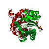
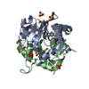
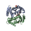
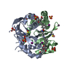
 Human immunodeficiency virus 1
 X-RAY DIFFRACTION /
  Authors
 Citation
 Structure visualization
 Downloads & links
 Other downloads


 PDBj
PDBj Assembly


 Mass: 96.063 Da / Num. of mol.: 4 / Source method: obtained synthetically / Formula: SO4
 Mass: 126.904 Da / Num. of mol.: 5 / Source method: obtained synthetically / Formula: I
 Mass: 334.322 Da / Num. of mol.: 1 / Source method: obtained synthetically / Formula: C20H14O5 / Feature type: SUBJECT OF INVESTIGATION
 Mass: 18.015 Da / Num. of mol.: 42 / Source method: isolated from a natural source / Formula: H2O
 Sample preparation
 / Beamline: MX2 / Wavelength: 0.954 Å
 Processing