| Entry | Database: PDB / ID: 6w0u
|
|---|
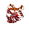
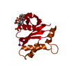
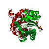
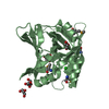
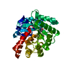
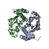
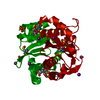
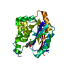
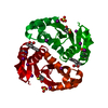
| Title | HIV Integrase core domain in complex with inhibitor 2-(2-ethynyl-5-methyl-1-benzofuran-3-yl)acetic acid |
|---|
 Components Components | Integrase |
|---|
 Keywords Keywords | TRANSFERASE/TRANSFERASE INHIBITOR / Inhibitor / HIV Integrase / TRANSFERASE-TRANSFERASE INHIBITOR complex |
|---|
| Function / homology |  Function and homology information Function and homology information
HIV-1 retropepsin / symbiont-mediated activation of host apoptosis / retroviral ribonuclease H / exoribonuclease H / exoribonuclease H activity / DNA integration / viral genome integration into host DNA / establishment of integrated proviral latency / RNA-directed DNA polymerase / RNA stem-loop binding ...HIV-1 retropepsin / symbiont-mediated activation of host apoptosis / retroviral ribonuclease H / exoribonuclease H / exoribonuclease H activity / DNA integration / viral genome integration into host DNA / establishment of integrated proviral latency / RNA-directed DNA polymerase / RNA stem-loop binding / viral penetration into host nucleus / host multivesicular body / RNA-directed DNA polymerase activity / RNA-DNA hybrid ribonuclease activity / Transferases; Transferring phosphorus-containing groups; Nucleotidyltransferases / host cell / viral nucleocapsid / endonuclease activity / DNA recombination / DNA-directed DNA polymerase / aspartic-type endopeptidase activity / Hydrolases; Acting on ester bonds / host cell cytoplasm / DNA-directed DNA polymerase activity / symbiont-mediated suppression of host gene expression / viral translational frameshifting / symbiont entry into host cell / lipid binding / host cell nucleus / host cell plasma membrane / virion membrane / structural molecule activity / proteolysis / DNA binding / zinc ion bindingSimilarity search - Function Reverse transcriptase connection / Reverse transcriptase connection domain / Reverse transcriptase thumb / Reverse transcriptase thumb domain / Integrase Zinc binding domain / Zinc finger integrase-type profile. / Integrase DNA binding domain / Integrase, C-terminal domain superfamily, retroviral / Integrase, N-terminal zinc-binding domain / Integrase-like, N-terminal ...Reverse transcriptase connection / Reverse transcriptase connection domain / Reverse transcriptase thumb / Reverse transcriptase thumb domain / Integrase Zinc binding domain / Zinc finger integrase-type profile. / Integrase DNA binding domain / Integrase, C-terminal domain superfamily, retroviral / Integrase, N-terminal zinc-binding domain / Integrase-like, N-terminal / Integrase, C-terminal, retroviral / Integrase DNA binding domain profile. / Immunodeficiency lentiviral matrix, N-terminal / gag gene protein p17 (matrix protein) / RNase H / Integrase core domain / Integrase, catalytic core / Integrase catalytic domain profile. / Retropepsin-like catalytic domain / Matrix protein, lentiviral and alpha-retroviral, N-terminal / RNase H type-1 domain profile. / Ribonuclease H domain / Retroviral nucleocapsid Gag protein p24, C-terminal domain / Gag protein p24 C-terminal domain / Retropepsins / Retroviral aspartyl protease / Aspartyl protease, retroviral-type family profile. / Peptidase A2A, retrovirus, catalytic / Retrovirus capsid, C-terminal / Reverse transcriptase (RNA-dependent DNA polymerase) / Retroviral matrix protein / Reverse transcriptase domain / Reverse transcriptase (RT) catalytic domain profile. / Retrovirus capsid, N-terminal / zinc finger / Zinc knuckle / Zinc finger, CCHC-type superfamily / Zinc finger, CCHC-type / Zinc finger CCHC-type profile. / Aspartic peptidase, active site / Eukaryotic and viral aspartyl proteases active site. / Aspartic peptidase domain superfamily / Ribonuclease H superfamily / Ribonuclease H-like superfamily / Reverse transcriptase/Diguanylate cyclase domain / DNA/RNA polymerase superfamilySimilarity search - Domain/homology |
|---|
| Biological species |   Human immunodeficiency virus 1 Human immunodeficiency virus 1 |
|---|
| Method |  X-RAY DIFFRACTION / FOURIER SYNTHESIS / Resolution: 2.19 Å X-RAY DIFFRACTION / FOURIER SYNTHESIS / Resolution: 2.19 Å |
|---|
 Authors Authors | Gorman, M.A. / Parker, M.W. |
|---|
 Citation Citation | Journal: To Be Published
Title: HIV Integrase core domain in complex with inhibitor
Authors: Gorman, M.A. / Parker, M.W. |
|---|
| History | | Deposition | Mar 2, 2020 | Deposition site: RCSB / Processing site: RCSB |
|---|
| Revision 1.0 | Mar 3, 2021 | Provider: repository / Type: Initial release |
|---|
| Revision 1.1 | Nov 13, 2024 | Group: Data collection / Database references / Structure summary
Category: chem_comp_atom / chem_comp_bond ...chem_comp_atom / chem_comp_bond / database_2 / pdbx_entry_details / pdbx_modification_feature
Item: _database_2.pdbx_DOI / _database_2.pdbx_database_accession / _pdbx_entry_details.has_protein_modification |
|---|
|
|---|
 Movie
Movie Controller

 Yorodumi
 Open data
 Basic information
 Structure visualization
 Downloads & links
 Other downloads
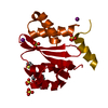


 PDBj
PDBj
 Assembly


 Mass: 214.217 Da / Num. of mol.: 1 / Source method: obtained synthetically / Formula: C13H10O3 / Feature type: SUBJECT OF INVESTIGATION
 Mass: 96.063 Da / Num. of mol.: 1 / Source method: obtained synthetically / Formula: SO4
 Mass: 126.904 Da / Num. of mol.: 5 / Source method: obtained synthetically / Formula: I
 Mass: 18.015 Da / Num. of mol.: 26 / Source method: isolated from a natural source / Formula: H2O
 Sample preparation
 Processing