 Movie
Movie Controller


+ Open data

- Basic information
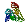
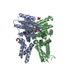
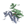
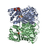
| Entry | Database: PDB / ID: 7l1e | ||||||||||||
|---|---|---|---|---|---|---|---|---|---|---|---|---|---|
| Title | The Crystal Structure of Bromide-Bound GtACR1 | ||||||||||||
 Components Components | Anion channelrhodopsin-1 | ||||||||||||
 Keywords Keywords | TRANSPORT PROTEIN / Bromide-Bound / Anion Tunnel / anion channel / rhodopsin | ||||||||||||
| Function / homology | Archaeal/bacterial/fungal rhodopsins / Bacteriorhodopsin-like protein / monoatomic ion channel activity / membrane => GO:0016020 / BROMIDE ION / (2R)-2,3-dihydroxypropyl (9Z)-octadec-9-enoate / Uncharacterized protein Function and homology information Function and homology information | ||||||||||||
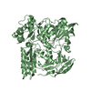
| Biological species |  | ||||||||||||
| Method |  X-RAY DIFFRACTION / SYNCHROTRON / MOLECULAR REPLACEMENT / Resolution: 3.2 Å X-RAY DIFFRACTION / SYNCHROTRON / MOLECULAR REPLACEMENT / Resolution: 3.2 Å | ||||||||||||
 Authors Authors | Li, H. / Huang, C.Y. / Wang, M. / Spudich, J.L. / Zheng, L. | ||||||||||||
| Funding support |  United States, United States,  Switzerland, 3items Switzerland, 3items
| ||||||||||||
 Citation Citation | Journal: Elife / Year: 2021 Title: The crystal structure of bromide-bound Gt ACR1 reveals a pre-activated state in the transmembrane anion tunnel. Authors: Li, H. / Huang, C.Y. / Govorunova, E.G. / Sineshchekov, O.A. / Yi, A. / Rothschild, K.J. / Wang, M. / Zheng, L. / Spudich, J.L. | ||||||||||||
| History |
|
- Structure visualization
| Structure viewer | Molecule: MolmilJmol/JSmol |
|---|
- Downloads & links
-Download
| PDBx/mmCIF format | 7l1e.cif.gz | 123.7 KB | Display | PDBx/mmCIF format |
|---|---|---|---|---|
| PDB format | pdb7l1e.ent.gz | 94.4 KB | Display | PDB format |
| PDBx/mmJSON format | 7l1e.json.gz | Tree view | PDBx/mmJSON format | |
| Others |  Other downloads Other downloads |
-Validation report
| Arichive directory | https://data.pdbj.org/pub/pdb/validation_reports/l1/7l1eftp://data.pdbj.org/pub/pdb/validation_reports/l1/7l1e | HTTPS FTP |
|---|
-Related structure data
| Related structure data | 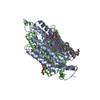 6edqS S: Starting model for refinement |
|---|---|
| Similar structure data |


-Links
 PDBj
PDBj


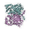
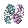
- Assembly
| Deposited unit | 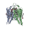
| ||||||||
|---|---|---|---|---|---|---|---|---|---|
| 1 |
| ||||||||
| Unit cell |
|
-Components
| #1: Protein | Mass: 30573.305 Da / Num. of mol.: 2 Source method: isolated from a genetically manipulated source Source: (gene. exp.)  Komagataella pastoris (fungus) / References: UniProt: A0A6U6DEE4 Komagataella pastoris (fungus) / References: UniProt: A0A6U6DEE4#2: Chemical |   Mass: 79.904 Da / Num. of mol.: 2 / Source method: obtained synthetically / Formula: Br / Feature type: SUBJECT OF INVESTIGATION Mass: 79.904 Da / Num. of mol.: 2 / Source method: obtained synthetically / Formula: Br / Feature type: SUBJECT OF INVESTIGATION#3: Chemical | ChemComp-OLC / (   Mass: 356.540 Da / Num. of mol.: 6 / Source method: obtained synthetically / Formula: C21H40O4 Mass: 356.540 Da / Num. of mol.: 6 / Source method: obtained synthetically / Formula: C21H40O4#4: Chemical |   Mass: 92.094 Da / Num. of mol.: 2 / Source method: obtained synthetically / Formula: C3H8O3 Mass: 92.094 Da / Num. of mol.: 2 / Source method: obtained synthetically / Formula: C3H8O3#5: Water | ChemComp-HOH / |  Mass: 18.015 Da / Num. of mol.: 17 / Source method: isolated from a natural source / Formula: H2O Mass: 18.015 Da / Num. of mol.: 17 / Source method: isolated from a natural source / Formula: H2OHas ligand of interest | Y | |
|---|
-Experimental details
-Experiment
| Experiment | Method: X-RAY DIFFRACTION / Number of used crystals: 1 |
|---|
- Sample preparation
| Crystal | Density Matthews: 2.53 Å3/Da / Density % sol: 51.38 % |
|---|---|
| Crystal grow | Temperature: 293 K / Method: lipidic cubic phase Details: 15% 2-methyl-2,4-pentanediol (MPD), 0.1 M NaBr with buffer 0.1 MES, pH 5.5 |
-Data collection
| Diffraction | Mean temperature: 100 K / Serial crystal experiment: Y |
|---|---|
| Diffraction source | Source: SYNCHROTRON / Site: SLS / Beamline: X06SA / Wavelength: 1 Å |
| Detector | Type: DECTRIS EIGER X 16M / Detector: PIXEL / Date: Sep 20, 2019 |
| Radiation | Protocol: SINGLE WAVELENGTH / Monochromatic (M) / Laue (L): M / Scattering type: x-ray |
| Radiation wavelength | Wavelength: 1 Å / Relative weight: 1 |
| Reflection | Resolution: 3.2→44.94 Å / Num. obs: 11033 / % possible obs: 95.6 % / Redundancy: 20 % / CC1/2: 0.975 / Net I/σ(I): 2.84 |
| Reflection shell | Resolution: 3.2→3.28 Å / Num. unique obs: 11033 / CC1/2: 0.975 |
| Serial crystallography sample delivery | Method: fixed target |
| Serial crystallography sample delivery fixed target | Description: COC film / Sample holding: IMISX h1 holder / Support base: IMISX h1 holder |
- Processing
| Software |
| |||||||||||||||||||||||||||||||||||
|---|---|---|---|---|---|---|---|---|---|---|---|---|---|---|---|---|---|---|---|---|---|---|---|---|---|---|---|---|---|---|---|---|---|---|---|---|
| Refinement | Method to determine structure: MOLECULAR REPLACEMENT Starting model: 6EDQ Resolution: 3.2→44.94 Å / SU ML: 0.58 / Cross valid method: THROUGHOUT / σ(F): 1.33 / Phase error: 29.87 / Stereochemistry target values: ML
| |||||||||||||||||||||||||||||||||||
| Solvent computation | Shrinkage radii: 0.9 Å / VDW probe radii: 1.11 Å / Solvent model: FLAT BULK SOLVENT MODEL | |||||||||||||||||||||||||||||||||||
| Refinement step | Cycle: LAST / Resolution: 3.2→44.94 Å
| |||||||||||||||||||||||||||||||||||
| Refine LS restraints |
| |||||||||||||||||||||||||||||||||||
| LS refinement shell |
|
