 Movie
Movie Controller

[English] 日本語
 Yorodumi
- PDB-3awj: Crystal structure of the Huperzia serrata polyketide synthase 1 c... -
+ Open data

- Basic information
| Entry | Database: PDB / ID: 3awj | ||||||
|---|---|---|---|---|---|---|---|
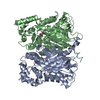
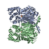
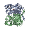
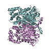
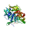
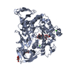
| Title | Crystal structure of the Huperzia serrata polyketide synthase 1 complexed with CoA-SH | ||||||
 Components Components | Chalcone synthase-like polyketide synthase | ||||||
 Keywords Keywords | TRANSFERASE / type III polyketide synthase / chalcone synthase | ||||||
| Function / homology |  Function and homology information Function and homology informationpolyketide biosynthetic process / acyltransferase activity, transferring groups other than amino-acyl groups Similarity search - Function | ||||||
| Biological species |  Huperzia serrata (toothed club-moss) Huperzia serrata (toothed club-moss) | ||||||
| Method |  X-RAY DIFFRACTION / SYNCHROTRON / MOLECULAR REPLACEMENT / Resolution: 2.2 Å X-RAY DIFFRACTION / SYNCHROTRON / MOLECULAR REPLACEMENT / Resolution: 2.2 Å | ||||||
 Authors Authors | Morita, H. / Kondo, S. / Kato, R. / Sugio, S. / Kohno, T. / Abe, I. | ||||||
 Citation Citation | Journal: Proc.Natl.Acad.Sci.USA / Year: 2011 Title: Synthesis of unnatural alkaloid scaffolds by exploiting plant polyketide synthase. Authors: Morita, H. / Yamashita, M. / Shi, S.P. / Wakimoto, T. / Kondo, S. / Kato, R. / Sugio, S. / Kohno, T. / Abe, I. | ||||||
| History |
|
- Structure visualization
| Structure viewer | Molecule: MolmilJmol/JSmol |
|---|
- Downloads & links
-Download
| PDBx/mmCIF format | 3awj.cif.gz | 159.4 KB | Display | PDBx/mmCIF format |
|---|---|---|---|---|
| PDB format | pdb3awj.ent.gz | 125.5 KB | Display | PDB format |
| PDBx/mmJSON format | 3awj.json.gz | Tree view | PDBx/mmJSON format | |
| Others |  Other downloads Other downloads |
-Validation report
| Arichive directory | https://data.pdbj.org/pub/pdb/validation_reports/aw/3awjftp://data.pdbj.org/pub/pdb/validation_reports/aw/3awj | HTTPS FTP |
|---|
-Related structure data
| Related structure data |  3awkC  1cgkS C: citing same article ( S: Starting model for refinement |
|---|---|
| Similar structure data |


-Links
 PDBj
PDBj


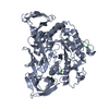
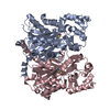
- Assembly
| Deposited unit | 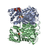
| ||||||||
|---|---|---|---|---|---|---|---|---|---|
| 1 |
| ||||||||
| Unit cell |
|
-Components
| #1: Protein | Mass: 43662.992 Da / Num. of mol.: 2 Source method: isolated from a genetically manipulated source Source: (gene. exp.) Huperzia serrata (toothed club-moss) / Gene: Pks1 / Plasmid: pET22b / Production host:  #2: Chemical | ChemComp-COA / |   Mass: 767.534 Da / Num. of mol.: 1 / Source method: obtained synthetically / Formula: C21H36N7O16P3S Mass: 767.534 Da / Num. of mol.: 1 / Source method: obtained synthetically / Formula: C21H36N7O16P3S#3: Chemical |   Mass: 96.063 Da / Num. of mol.: 2 / Source method: obtained synthetically / Formula: SO4 Mass: 96.063 Da / Num. of mol.: 2 / Source method: obtained synthetically / Formula: SO4#4: Water | ChemComp-HOH / |  Mass: 18.015 Da / Num. of mol.: 227 / Source method: isolated from a natural source / Formula: H2O Mass: 18.015 Da / Num. of mol.: 227 / Source method: isolated from a natural source / Formula: H2OHas protein modification | Y | |
|---|
-Experimental details
-Experiment
| Experiment | Method: X-RAY DIFFRACTION / Number of used crystals: 1 |
|---|
- Sample preparation
| Crystal | Density Matthews: 2.51 Å3/Da / Density % sol: 50.92 % |
|---|---|
| Crystal grow | Temperature: 293 K / Method: vapor diffusion, hanging drop / pH: 6.5 Details: 0.1M MES-NaOH, 1925mM ammonium sulfate, 1,1,1,3,3,3-hexafluoro-2-propanol, 2mM CoA-SH, pH 6.5, VAPOR DIFFUSION, HANGING DROP, temperature 293K |
-Data collection
| Diffraction | Mean temperature: 100 K |
|---|---|
| Diffraction source | Source: SYNCHROTRON / Site: SPring-8  / Beamline: BL24XU / Wavelength: 0.82656 Å / Beamline: BL24XU / Wavelength: 0.82656 Å |
| Detector | Type: RIGAKU RAXIS V / Detector: IMAGE PLATE / Date: Apr 16, 2007 |
| Radiation | Monochromator: bending magnet / Protocol: SINGLE WAVELENGTH / Monochromatic (M) / Laue (L): M / Scattering type: x-ray |
| Radiation wavelength | Wavelength: 0.82656 Å / Relative weight: 1 |
| Reflection | Resolution: 2.2→30 Å / Num. all: 42570 / Num. obs: 42570 / % possible obs: 99.7 % / Observed criterion σ(F): 0 / Observed criterion σ(I): 0 / Redundancy: 6.6 % / Rsym value: 0.092 / Net I/σ(I): 34.3 |
| Reflection shell | Resolution: 2.2→2.28 Å / Redundancy: 5.9 % / Mean I/σ(I) obs: 9.2 / Num. unique all: 4368 / Rsym value: 0.288 / % possible all: 98.3 |
- Processing
| Software |
| ||||||||||||||||||||
|---|---|---|---|---|---|---|---|---|---|---|---|---|---|---|---|---|---|---|---|---|---|
| Refinement | Method to determine structure: MOLECULAR REPLACEMENT Starting model: 1CGK Resolution: 2.2→30 Å / σ(F): 0 / Stereochemistry target values: Engh & Huber
| ||||||||||||||||||||
| Refinement step | Cycle: LAST / Resolution: 2.2→30 Å
| ||||||||||||||||||||
| Refine LS restraints |
|
