 Movie
Movie Controller

[English] 日本語
 Yorodumi
- PDB-7kwx: Spermidine N-acetyltransferase SpeG N152L mutant from Vibrio cholerae -
+ Open data

- Basic information
| Entry | Database: PDB / ID: 7kwx | ||||||
|---|---|---|---|---|---|---|---|
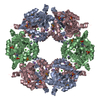
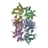
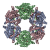
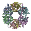
| Title | Spermidine N-acetyltransferase SpeG N152L mutant from Vibrio cholerae | ||||||
 Components Components | Spermidine N(1)-acetyltransferase | ||||||
 Keywords Keywords | TRANSFERASE / SpeG Enzyme Allosteric | ||||||
| Function / homology |  Function and homology information Function and homology informationpolyamine catabolic process / spermine catabolic process / diamine N-acetyltransferase / diamine N-acetyltransferase activity / spermidine catabolic process / magnesium ion binding / cytoplasm Similarity search - Function | ||||||
| Biological species |  Vibrio cholerae serotype O1 (bacteria) Vibrio cholerae serotype O1 (bacteria) | ||||||
| Method |  X-RAY DIFFRACTION / SYNCHROTRON / MOLECULAR REPLACEMENT / Resolution: 2.42 Å X-RAY DIFFRACTION / SYNCHROTRON / MOLECULAR REPLACEMENT / Resolution: 2.42 Å | ||||||
 Authors Authors | Le, V.T.B. / Tsimbalyuk, S. / Lim, E.Q. / Solis, A. / Gawat, D. / Boeck, P. / Renolo, R. / Forwood, J.K. / Kuhn, M.L. | ||||||
 Citation Citation | Journal: Front Mol Biosci / Year: 2021 Title: The Vibrio cholerae SpeG Spermidine/Spermine N -Acetyltransferase Allosteric Loop and beta 6-beta 7 Structural Elements Are Critical for Kinetic Activity. Authors: Le, V.T.B. / Tsimbalyuk, S. / Lim, E.Q. / Solis, A. / Gawat, D. / Boeck, P. / Lim, E.Q. / Renolo, R. / Forwood, J.K. / Kuhn, M.L. | ||||||
| History |
|
- Structure visualization
| Structure viewer | Molecule: MolmilJmol/JSmol |
|---|
- Downloads & links
-Download
| PDBx/mmCIF format | 7kwx.cif.gz | 250.6 KB | Display | PDBx/mmCIF format |
|---|---|---|---|---|
| PDB format | pdb7kwx.ent.gz | 166.8 KB | Display | PDB format |
| PDBx/mmJSON format | 7kwx.json.gz | Tree view | PDBx/mmJSON format | |
| Others |  Other downloads Other downloads |
-Validation report
| Arichive directory | https://data.pdbj.org/pub/pdb/validation_reports/kw/7kwxftp://data.pdbj.org/pub/pdb/validation_reports/kw/7kwx | HTTPS FTP |
|---|
-Related structure data
| Related structure data |  7kwhC 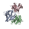 7kwjC 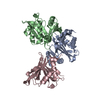 7kwqC 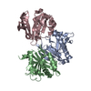 7kx2C 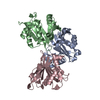 7kx3C 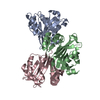 4jjxS S: Starting model for refinement C: citing same article ( |
|---|---|
| Similar structure data |


-Links
 PDBj
PDBj

- Assembly
| Deposited unit | 
| ||||||||||||
|---|---|---|---|---|---|---|---|---|---|---|---|---|---|
| 1 | 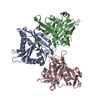
| ||||||||||||
| Unit cell |
|
-Components
| #1: Protein | Mass: 20702.441 Da / Num. of mol.: 3 / Mutation: N152L Source method: isolated from a genetically manipulated source Source: (gene. exp.) Vibrio cholerae serotype O1 (strain ATCC 39315 / El Tor Inaba N16961) (bacteria)Strain: ATCC 39315 / El Tor Inaba N16961 / Gene: speG, VC_A0947 / Production host: |
|---|
-Experimental details
-Experiment
| Experiment | Method: X-RAY DIFFRACTION / Number of used crystals: 1 |
|---|
- Sample preparation
| Crystal | Density Matthews: 2.78 Å3/Da / Density % sol: 55.83 % |
|---|---|
| Crystal grow | Temperature: 295 K / Method: vapor diffusion, hanging drop / Details: 10% ethanol |
-Data collection
| Diffraction | Mean temperature: 100 K / Serial crystal experiment: N |
|---|---|
| Diffraction source | Source: SYNCHROTRON / Site: Australian Synchrotron  / Beamline: MX1 / Wavelength: 0.9537 Å / Beamline: MX1 / Wavelength: 0.9537 Å |
| Detector | Type: DECTRIS EIGER X 16M / Detector: PIXEL / Date: Apr 7, 2018 |
| Radiation | Protocol: SINGLE WAVELENGTH / Monochromatic (M) / Laue (L): M / Scattering type: x-ray |
| Radiation wavelength | Wavelength: 0.9537 Å / Relative weight: 1 |
| Reflection | Resolution: 2.42→48.66 Å / Num. obs: 26869 / % possible obs: 100 % / Redundancy: 8.7 % / Biso Wilson estimate: 46.32 Å2 / CC1/2: 0.998 / Net I/σ(I): 16.1 |
| Reflection shell | Resolution: 2.42→2.507 Å / Mean I/σ(I) obs: 2.4 / Num. unique obs: 2762 / CC1/2: 0.778 |
- Processing
| Software |
| |||||||||||||||||||||||||||||||||||||||||||||||||||||||||||||||||||||||||||||
|---|---|---|---|---|---|---|---|---|---|---|---|---|---|---|---|---|---|---|---|---|---|---|---|---|---|---|---|---|---|---|---|---|---|---|---|---|---|---|---|---|---|---|---|---|---|---|---|---|---|---|---|---|---|---|---|---|---|---|---|---|---|---|---|---|---|---|---|---|---|---|---|---|---|---|---|---|---|---|
| Refinement | Method to determine structure: MOLECULAR REPLACEMENT Starting model: 4JJX Resolution: 2.42→47.27 Å / SU ML: 0.3573 / Cross valid method: FREE R-VALUE / σ(F): 1.33 / Phase error: 29.7465 Stereochemistry target values: GeoStd + Monomer Library + CDL v1.2
| |||||||||||||||||||||||||||||||||||||||||||||||||||||||||||||||||||||||||||||
| Solvent computation | Shrinkage radii: 0.9 Å / VDW probe radii: 1.11 Å / Solvent model: FLAT BULK SOLVENT MODEL | |||||||||||||||||||||||||||||||||||||||||||||||||||||||||||||||||||||||||||||
| Displacement parameters | Biso mean: 52.16 Å2 | |||||||||||||||||||||||||||||||||||||||||||||||||||||||||||||||||||||||||||||
| Refinement step | Cycle: LAST / Resolution: 2.42→47.27 Å
| |||||||||||||||||||||||||||||||||||||||||||||||||||||||||||||||||||||||||||||
| Refine LS restraints |
| |||||||||||||||||||||||||||||||||||||||||||||||||||||||||||||||||||||||||||||
| LS refinement shell |
|
