[English] 日本語
 Yorodumi
- PDB-7kuy: Cyro-EM structure of human Glycine Receptor alpha2-beta heteromer... -
+ Open data
- Basic information
| Entry | Database: PDB / ID: 7kuy | ||||||
|---|---|---|---|---|---|---|---|
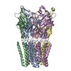
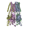
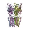
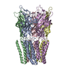
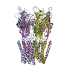
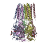
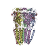
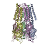
| Title | Cyro-EM structure of human Glycine Receptor alpha2-beta heteromer, strychnine bound state | ||||||
 Components Components |
| ||||||
 Keywords Keywords | MEMBRANE PROTEIN / SIGNALING PROTEIN / glycine receptor / alpha2-beta hetero-pentamer / strychnine | ||||||
| Function / homology |  Function and homology information Function and homology informationglycine-gated chloride ion channel activity / acrosome reaction / synaptic transmission, glycinergic / glycine-gated chloride channel complex / Neurotransmitter receptors and postsynaptic signal transmission / gamma-aminobutyric acid receptor clustering / postsynaptic specialization / extracellularly glycine-gated ion channel activity / righting reflex / extracellularly glycine-gated chloride channel activity ...glycine-gated chloride ion channel activity / acrosome reaction / synaptic transmission, glycinergic / glycine-gated chloride channel complex / Neurotransmitter receptors and postsynaptic signal transmission / gamma-aminobutyric acid receptor clustering / postsynaptic specialization / extracellularly glycine-gated ion channel activity / righting reflex / extracellularly glycine-gated chloride channel activity / glycinergic synapse / adult walking behavior / postsynaptic specialization membrane / glycine binding / cellular response to zinc ion / startle response / cellular response to ethanol / chloride channel complex / neuropeptide signaling pathway / monoatomic ion transport / visual perception / chloride transmembrane transport / bioluminescence / cell projection / generation of precursor metabolites and energy / transmitter-gated monoatomic ion channel activity involved in regulation of postsynaptic membrane potential / cellular response to amino acid stimulus / modulation of chemical synaptic transmission / GABA-ergic synapse / transmembrane signaling receptor activity / nervous system development / monoatomic ion transmembrane transport / chemical synaptic transmission / postsynaptic membrane / intracellular membrane-bounded organelle / dendrite / protein-containing complex binding / metal ion binding / plasma membrane / cytoplasm Similarity search - Function | ||||||
| Biological species |  Homo sapiens (human) Homo sapiens (human) | ||||||
| Method | ELECTRON MICROSCOPY / single particle reconstruction / cryo EM / Resolution: 3.6 Å | ||||||
 Authors Authors | Yu, H. / Wang, W. | ||||||
| Funding support |  United States, 1items United States, 1items
| ||||||
 Citation Citation |  Journal: Neuron / Year: 2021 Journal: Neuron / Year: 2021Title: Characterization of the subunit composition and structure of adult human glycine receptors. Authors: Hailong Yu / Xiao-Chen Bai / Weiwei Wang /  Abstract: The strychnine-sensitive pentameric glycine receptor (GlyR) mediates fast inhibitory neurotransmission in the mammalian nervous system. Only heteromeric GlyRs mediate synaptic transmission, as they ...The strychnine-sensitive pentameric glycine receptor (GlyR) mediates fast inhibitory neurotransmission in the mammalian nervous system. Only heteromeric GlyRs mediate synaptic transmission, as they contain the β subunit that permits clustering at the synapse through its interaction with scaffolding proteins. Here, we show that α2 and β subunits assemble with an unexpected 4:1 stoichiometry to produce GlyR with native electrophysiological properties. We determined structures in multiple functional states at 3.6-3.8 Å resolutions and show how 4:1 stoichiometry is consistent with the structural features of α2β GlyR. Furthermore, we show that one single β subunit in each GlyR gives rise to the characteristic electrophysiological properties of heteromeric GlyR, while more β subunits render GlyR non-conductive. A single β subunit ensures a univalent GlyR-scaffold linkage, which means the scaffold alone regulates the cluster properties. | ||||||
| History |
|
- Structure visualization
| Movie |
 Movie viewer Movie viewer |
|---|---|
| Structure viewer | Molecule:  Molmil Molmil Jmol/JSmol Jmol/JSmol |
- Downloads & links
- Download
| PDBx/mmCIF format |  7kuy.cif.gz 7kuy.cif.gz | 316.7 KB | Display |  PDBx/mmCIF format PDBx/mmCIF format |
|---|---|---|---|---|
| PDB format |  pdb7kuy.ent.gz pdb7kuy.ent.gz | 251.5 KB | Display |  PDB format PDB format |
| PDBx/mmJSON format |  7kuy.json.gz 7kuy.json.gz | Tree view |  PDBx/mmJSON format PDBx/mmJSON format | |
| Others |  Other downloads Other downloads |
-Validation report
| Arichive directory |  https://data.pdbj.org/pub/pdb/validation_reports/ku/7kuy https://data.pdbj.org/pub/pdb/validation_reports/ku/7kuy ftp://data.pdbj.org/pub/pdb/validation_reports/ku/7kuy ftp://data.pdbj.org/pub/pdb/validation_reports/ku/7kuy | HTTPS FTP |
|---|
-Related structure data
| Related structure data |  23041MC  9403C  9404C  5bkfC  5bkgC  7l31C M: map data used to model this data C: citing same article ( |
|---|---|
| Similar structure data |
- Links
Links
- Assembly
| Deposited unit | 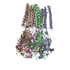
|
|---|---|
| 1 |
|
- Components
| #1: Protein | Mass: 41676.004 Da / Num. of mol.: 4 Source method: isolated from a genetically manipulated source Source: (gene. exp.)  Homo sapiens (human) / Gene: GLRA2 / Production host: Homo sapiens (human) / Gene: GLRA2 / Production host:  Homo sapiens (human) / References: UniProt: P23416 Homo sapiens (human) / References: UniProt: P23416#2: Protein | | Mass: 78765.086 Da / Num. of mol.: 1 Source method: isolated from a genetically manipulated source Source: (gene. exp.)  Homo sapiens (human), (gene. exp.) Homo sapiens (human), (gene. exp.)  Gene: GLRB, GFP / Production host:  Homo sapiens (human) / References: UniProt: P48167, UniProt: P42212 Homo sapiens (human) / References: UniProt: P48167, UniProt: P42212#3: Sugar | ChemComp-NAG / #4: Chemical | ChemComp-SY9 / Has ligand of interest | Y | Has protein modification | Y | |
|---|
-Experimental details
-Experiment
| Experiment | Method: ELECTRON MICROSCOPY |
|---|---|
| EM experiment | Aggregation state: PARTICLE / 3D reconstruction method: single particle reconstruction |
- Sample preparation
| Component |
| ||||||||||||||||||||||||
|---|---|---|---|---|---|---|---|---|---|---|---|---|---|---|---|---|---|---|---|---|---|---|---|---|---|
| Molecular weight | Value: 0.257 MDa / Experimental value: YES | ||||||||||||||||||||||||
| Source (natural) |
| ||||||||||||||||||||||||
| Source (recombinant) |
| ||||||||||||||||||||||||
| Buffer solution | pH: 8 | ||||||||||||||||||||||||
| Buffer component |
| ||||||||||||||||||||||||
| Specimen | Conc.: 3.5 mg/ml / Embedding applied: NO / Shadowing applied: NO / Staining applied: NO / Vitrification applied: YES | ||||||||||||||||||||||||
| Specimen support | Grid material: GOLD / Grid mesh size: 400 divisions/in. / Grid type: Quantifoil R1.2/1.3 | ||||||||||||||||||||||||
| Vitrification | Instrument: FEI VITROBOT MARK IV / Cryogen name: ETHANE / Humidity: 100 % / Chamber temperature: 298 K |
- Electron microscopy imaging
| Experimental equipment |  Model: Titan Krios / Image courtesy: FEI Company |
|---|---|
| Microscopy | Model: FEI TITAN KRIOS |
| Electron gun | Electron source:  FIELD EMISSION GUN / Accelerating voltage: 300 kV / Illumination mode: FLOOD BEAM FIELD EMISSION GUN / Accelerating voltage: 300 kV / Illumination mode: FLOOD BEAM |
| Electron lens | Mode: BRIGHT FIELD / Nominal magnification: 105000 X / Nominal defocus max: 2500 nm / Nominal defocus min: 1000 nm / Calibrated defocus min: 800 nm / Calibrated defocus max: 3200 nm / Alignment procedure: BASIC |
| Specimen holder | Cryogen: NITROGEN / Specimen holder model: FEI TITAN KRIOS AUTOGRID HOLDER |
| Image recording | Electron dose: 51 e/Å2 / Film or detector model: GATAN K3 (6k x 4k) / Num. of grids imaged: 1 / Num. of real images: 4111 |
| EM imaging optics | Energyfilter name: GIF Quantum LS / Energyfilter slit width: 20 eV |
- Processing
| EM software |
| ||||||||||||||||||||||||||||||
|---|---|---|---|---|---|---|---|---|---|---|---|---|---|---|---|---|---|---|---|---|---|---|---|---|---|---|---|---|---|---|---|
| CTF correction | Type: PHASE FLIPPING AND AMPLITUDE CORRECTION | ||||||||||||||||||||||||||||||
| Particle selection | Num. of particles selected: 472972 | ||||||||||||||||||||||||||||||
| Symmetry | Point symmetry: C1 (asymmetric) | ||||||||||||||||||||||||||||||
| 3D reconstruction | Resolution: 3.6 Å / Resolution method: FSC 0.143 CUT-OFF / Num. of particles: 44965 / Symmetry type: POINT | ||||||||||||||||||||||||||||||
| Atomic model building | Space: REAL | ||||||||||||||||||||||||||||||
| Atomic model building | PDB-ID: 3JAD Accession code: 3JAD / Source name: PDB / Type: experimental model |
 Movie
Movie Controller


 PDBj
PDBj


