[English] 日本語
 Yorodumi
- EMDB-23148: Cyro-EM structure of human Glycine Receptor alpha2-beta heteromer... -
+ Open data
- Basic information
| Entry | Database: EMDB / ID: EMD-23148 | |||||||||
|---|---|---|---|---|---|---|---|---|---|---|
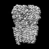
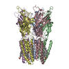
| Title | Cyro-EM structure of human Glycine Receptor alpha2-beta heteromer, strychnine bound state, 3.8 Angstrom | |||||||||
 Map data Map data | ||||||||||
 Sample Sample |
| |||||||||
 Keywords Keywords | glycine receptor / alpha2-beta hetero-pentamer / strychnine / MEMBRANE PROTEIN | |||||||||
| Function / homology |  Function and homology information Function and homology informationglycine-gated chloride ion channel activity / acrosome reaction / synaptic transmission, glycinergic / glycine-gated chloride channel complex / Neurotransmitter receptors and postsynaptic signal transmission / gamma-aminobutyric acid receptor clustering / postsynaptic specialization / extracellularly glycine-gated ion channel activity / righting reflex / extracellularly glycine-gated chloride channel activity ...glycine-gated chloride ion channel activity / acrosome reaction / synaptic transmission, glycinergic / glycine-gated chloride channel complex / Neurotransmitter receptors and postsynaptic signal transmission / gamma-aminobutyric acid receptor clustering / postsynaptic specialization / extracellularly glycine-gated ion channel activity / righting reflex / extracellularly glycine-gated chloride channel activity / glycinergic synapse / adult walking behavior / postsynaptic specialization membrane / glycine binding / cellular response to zinc ion / startle response / cellular response to ethanol / chloride channel complex / neuropeptide signaling pathway / monoatomic ion transport / visual perception / chloride transmembrane transport / bioluminescence / generation of precursor metabolites and energy / cell projection / transmitter-gated monoatomic ion channel activity involved in regulation of postsynaptic membrane potential / cellular response to amino acid stimulus / modulation of chemical synaptic transmission / GABA-ergic synapse / transmembrane signaling receptor activity / nervous system development / monoatomic ion transmembrane transport / chemical synaptic transmission / postsynaptic membrane / intracellular membrane-bounded organelle / dendrite / protein-containing complex binding / metal ion binding / plasma membrane / cytoplasm Similarity search - Function | |||||||||
| Biological species |  Homo sapiens (human) Homo sapiens (human) | |||||||||
| Method | single particle reconstruction / cryo EM / Resolution: 3.8 Å | |||||||||
 Authors Authors | Yu H / Wang W | |||||||||
| Funding support |  United States, 1 items United States, 1 items
| |||||||||
 Citation Citation |  Journal: Neuron / Year: 2021 Journal: Neuron / Year: 2021Title: Characterization of the subunit composition and structure of adult human glycine receptors. Authors: Hailong Yu / Xiao-Chen Bai / Weiwei Wang /  Abstract: The strychnine-sensitive pentameric glycine receptor (GlyR) mediates fast inhibitory neurotransmission in the mammalian nervous system. Only heteromeric GlyRs mediate synaptic transmission, as they ...The strychnine-sensitive pentameric glycine receptor (GlyR) mediates fast inhibitory neurotransmission in the mammalian nervous system. Only heteromeric GlyRs mediate synaptic transmission, as they contain the β subunit that permits clustering at the synapse through its interaction with scaffolding proteins. Here, we show that α2 and β subunits assemble with an unexpected 4:1 stoichiometry to produce GlyR with native electrophysiological properties. We determined structures in multiple functional states at 3.6-3.8 Å resolutions and show how 4:1 stoichiometry is consistent with the structural features of α2β GlyR. Furthermore, we show that one single β subunit in each GlyR gives rise to the characteristic electrophysiological properties of heteromeric GlyR, while more β subunits render GlyR non-conductive. A single β subunit ensures a univalent GlyR-scaffold linkage, which means the scaffold alone regulates the cluster properties. | |||||||||
| History |
|
- Structure visualization
| Movie |
 Movie viewer Movie viewer |
|---|---|
| Structure viewer | EM map:  SurfView SurfView Molmil Molmil Jmol/JSmol Jmol/JSmol |
| Supplemental images |
- Downloads & links
-EMDB archive
| Map data |  emd_23148.map.gz emd_23148.map.gz | 59 MB |  EMDB map data format EMDB map data format | |
|---|---|---|---|---|
| Header (meta data) |  emd-23148-v30.xml emd-23148-v30.xml emd-23148.xml emd-23148.xml | 17.1 KB 17.1 KB | Display Display |  EMDB header EMDB header |
| Images |  emd_23148.png emd_23148.png | 134.3 KB | ||
| Filedesc metadata |  emd-23148.cif.gz emd-23148.cif.gz | 6.7 KB | ||
| Archive directory |  http://ftp.pdbj.org/pub/emdb/structures/EMD-23148 http://ftp.pdbj.org/pub/emdb/structures/EMD-23148 ftp://ftp.pdbj.org/pub/emdb/structures/EMD-23148 ftp://ftp.pdbj.org/pub/emdb/structures/EMD-23148 | HTTPS FTP |
-Validation report
| Summary document |  emd_23148_validation.pdf.gz emd_23148_validation.pdf.gz | 558.9 KB | Display |  EMDB validaton report EMDB validaton report |
|---|---|---|---|---|
| Full document |  emd_23148_full_validation.pdf.gz emd_23148_full_validation.pdf.gz | 558.4 KB | Display | |
| Data in XML |  emd_23148_validation.xml.gz emd_23148_validation.xml.gz | 6 KB | Display | |
| Data in CIF |  emd_23148_validation.cif.gz emd_23148_validation.cif.gz | 6.8 KB | Display | |
| Arichive directory |  https://ftp.pdbj.org/pub/emdb/validation_reports/EMD-23148 https://ftp.pdbj.org/pub/emdb/validation_reports/EMD-23148 ftp://ftp.pdbj.org/pub/emdb/validation_reports/EMD-23148 ftp://ftp.pdbj.org/pub/emdb/validation_reports/EMD-23148 | HTTPS FTP |
-Related structure data
| Related structure data |  7l31MC  9403C  9404C  5bkfC  5bkgC  7kuyC M: atomic model generated by this map C: citing same article ( |
|---|---|
| Similar structure data |
- Links
Links
| EMDB pages |  EMDB (EBI/PDBe) / EMDB (EBI/PDBe) /  EMDataResource EMDataResource |
|---|---|
| Related items in Molecule of the Month |
- Map
Map
| File |  Download / File: emd_23148.map.gz / Format: CCP4 / Size: 64 MB / Type: IMAGE STORED AS FLOATING POINT NUMBER (4 BYTES) Download / File: emd_23148.map.gz / Format: CCP4 / Size: 64 MB / Type: IMAGE STORED AS FLOATING POINT NUMBER (4 BYTES) | ||||||||||||||||||||||||||||||||||||||||||||||||||||||||||||||||||||
|---|---|---|---|---|---|---|---|---|---|---|---|---|---|---|---|---|---|---|---|---|---|---|---|---|---|---|---|---|---|---|---|---|---|---|---|---|---|---|---|---|---|---|---|---|---|---|---|---|---|---|---|---|---|---|---|---|---|---|---|---|---|---|---|---|---|---|---|---|---|
| Projections & slices | Image control
Images are generated by Spider. | ||||||||||||||||||||||||||||||||||||||||||||||||||||||||||||||||||||
| Voxel size | X=Y=Z: 0.83 Å | ||||||||||||||||||||||||||||||||||||||||||||||||||||||||||||||||||||
| Density |
| ||||||||||||||||||||||||||||||||||||||||||||||||||||||||||||||||||||
| Symmetry | Space group: 1 | ||||||||||||||||||||||||||||||||||||||||||||||||||||||||||||||||||||
| Details | EMDB XML:
CCP4 map header:
| ||||||||||||||||||||||||||||||||||||||||||||||||||||||||||||||||||||
-Supplemental data
- Sample components
-Entire : Glycine receptor alpha2-beta heteromer with strychnine, 3.8 Angstrom
| Entire | Name: Glycine receptor alpha2-beta heteromer with strychnine, 3.8 Angstrom |
|---|---|
| Components |
|
-Supramolecule #1: Glycine receptor alpha2-beta heteromer with strychnine, 3.8 Angstrom
| Supramolecule | Name: Glycine receptor alpha2-beta heteromer with strychnine, 3.8 Angstrom type: complex / ID: 1 / Parent: 0 / Macromolecule list: #1-#2 |
|---|---|
| Molecular weight | Theoretical: 257 KDa |
-Supramolecule #2: Glycine receptor subunit alpha-2
| Supramolecule | Name: Glycine receptor subunit alpha-2 / type: organelle_or_cellular_component / ID: 2 / Parent: 1 / Macromolecule list: #1 |
|---|---|
| Source (natural) | Organism:  Homo sapiens (human) Homo sapiens (human) |
-Supramolecule #3: Glycine receptor subunit beta, Green fluorescent protein chimera
| Supramolecule | Name: Glycine receptor subunit beta, Green fluorescent protein chimera type: organelle_or_cellular_component / ID: 3 / Parent: 1 / Macromolecule list: #2 |
|---|---|
| Source (natural) | Organism:  Homo sapiens (human) Homo sapiens (human) |
-Macromolecule #1: Glycine receptor subunit alpha-2
| Macromolecule | Name: Glycine receptor subunit alpha-2 / type: protein_or_peptide / ID: 1 / Number of copies: 4 / Enantiomer: LEVO |
|---|---|
| Source (natural) | Organism:  Homo sapiens (human) Homo sapiens (human) |
| Molecular weight | Theoretical: 41.676004 KDa |
| Recombinant expression | Organism:  Homo sapiens (human) Homo sapiens (human) |
| Sequence | String: KDHDSRSGKQ PSQTLSPSDF LDKLMGRTSG YDARIRPNFK GPPVNVTCNI FINSFGSVTE TTMDYRVNIF LRQQWNDSRL AYSEYPDDS LDLDPSMLDS IWKPDLFFAN EKGANFHDVT TDNKLLRISK NGKVLYSIRL TLTLSCPMDL KNFPMDVQTC T MQLESFGY ...String: KDHDSRSGKQ PSQTLSPSDF LDKLMGRTSG YDARIRPNFK GPPVNVTCNI FINSFGSVTE TTMDYRVNIF LRQQWNDSRL AYSEYPDDS LDLDPSMLDS IWKPDLFFAN EKGANFHDVT TDNKLLRISK NGKVLYSIRL TLTLSCPMDL KNFPMDVQTC T MQLESFGY TMNDLIFEWL SDGPVQVAEG LTLPQFILKE EKELGYCTKH YNTGKFTCIE VKFHLERQMG YYLIQMYIPS LL IVILSWV SFWINMDAAP ARVALGITTV LTMTTQSSGS RASLPKVSYV KAIDIWMAVC LLFVFAALLE YAAVNFVSRG SSG KKFVDR AKRIDTISRA AFPLAFLIFN IFYWITYKII RHEDVHKK UniProtKB: Glycine receptor subunit alpha-2, Glycine receptor subunit alpha-2 |
-Macromolecule #2: Glycine receptor subunit beta,Green fluorescent protein
| Macromolecule | Name: Glycine receptor subunit beta,Green fluorescent protein type: protein_or_peptide / ID: 2 Details: This is a GFP insertion between Glycine Receptor Beta M3-M4 helix Number of copies: 1 / Enantiomer: LEVO |
|---|---|
| Source (natural) | Organism:  Homo sapiens (human) Homo sapiens (human) |
| Molecular weight | Theoretical: 78.765086 KDa |
| Recombinant expression | Organism:  Homo sapiens (human) Homo sapiens (human) |
| Sequence | String: GVAMPGAEDD VVAALEVLFQ GPKSSKKGKG KKKQYLCPSQ QSAEDLARVP ANSTSNILNR LLVSYDPRIR PNFKGIPVDV VVNIFINSF GSIQETTMDY RVNIFLRQKW NDPRLKLPSD FRGSDALTVD PTMYKCLWKP DLFFANEKSA NFHDVTQENI L LFIFRDGD ...String: GVAMPGAEDD VVAALEVLFQ GPKSSKKGKG KKKQYLCPSQ QSAEDLARVP ANSTSNILNR LLVSYDPRIR PNFKGIPVDV VVNIFINSF GSIQETTMDY RVNIFLRQKW NDPRLKLPSD FRGSDALTVD PTMYKCLWKP DLFFANEKSA NFHDVTQENI L LFIFRDGD VLVSMRLSIT LSCPLDLTLF PMDTQRCKMQ LESFGYTTDD LRFIWQSGDP VQLEKIALPQ FDIKKEDIEY GN CTKYYKG TGYYTCVEVI FTLRRQVGFY MMGVYAPTLL IVVLSWLSFW INPDASAARV PLGIFSVLSL ASECTTLAAE LPK VSYVKA LDVWLIACLL FGFASLVEYA VVQVMLNGGS SAAAVSKGEE LFTGVVPILV ELDGDVNGHK FSVSGEGEGD ATYG KLTLK FICTTGKLPV PWPTLVTTLT YGVQCFSRYP DHMKQHDFFK SAMPEGYVQE RTIFFKDDGN YKTRAEVKFE GDTLV NRIE LKGIDFKEDG NILGHKLEYN YNSHNVYIMA DKQKNGIKVN FKIRHNIEDG SVQLADHYQQ NTPIGDGPVL LPDNHY LST QSKLSKDPNE KRDHMVLLEF VTAAGITLGM DELYKSGSGS GVGETRCKKV CTSKSDLRSN DFSIVGSLPR DFELSNY DC YGKPIEVNNG LGKSQAKNNK KPPPAKPVIP TAAKRIDLYA RALFPFCFLF FNVIYWSIYL UniProtKB: Glycine receptor subunit beta, Green fluorescent protein, Glycine receptor subunit beta |
-Macromolecule #3: 2-acetamido-2-deoxy-beta-D-glucopyranose
| Macromolecule | Name: 2-acetamido-2-deoxy-beta-D-glucopyranose / type: ligand / ID: 3 / Number of copies: 9 / Formula: NAG |
|---|---|
| Molecular weight | Theoretical: 221.208 Da |
| Chemical component information |  ChemComp-NAG: |
-Macromolecule #4: STRYCHNINE
| Macromolecule | Name: STRYCHNINE / type: ligand / ID: 4 / Number of copies: 5 / Formula: SY9 |
|---|---|
| Molecular weight | Theoretical: 334.412 Da |
| Chemical component information |  ChemComp-SY9: |
-Experimental details
-Structure determination
| Method | cryo EM |
|---|---|
 Processing Processing | single particle reconstruction |
| Aggregation state | particle |
- Sample preparation
| Concentration | 3.5 mg/mL | ||||||||
|---|---|---|---|---|---|---|---|---|---|
| Buffer | pH: 8 / Component:
| ||||||||
| Grid | Model: Quantifoil R1.2/1.3 / Material: GOLD / Mesh: 400 / Support film - Material: CARBON / Support film - topology: HOLEY / Pretreatment - Type: GLOW DISCHARGE / Pretreatment - Time: 30 sec. / Pretreatment - Atmosphere: AIR / Pretreatment - Pressure: 0.038 kPa | ||||||||
| Vitrification | Cryogen name: ETHANE / Chamber humidity: 100 % / Chamber temperature: 298 K / Instrument: FEI VITROBOT MARK IV |
- Electron microscopy
| Microscope | FEI TITAN KRIOS |
|---|---|
| Specialist optics | Energy filter - Name: GIF Quantum LS / Energy filter - Slit width: 20 eV |
| Image recording | Film or detector model: GATAN K3 (6k x 4k) / Number grids imaged: 1 / Number real images: 4111 / Average electron dose: 51.0 e/Å2 |
| Electron beam | Acceleration voltage: 300 kV / Electron source:  FIELD EMISSION GUN FIELD EMISSION GUN |
| Electron optics | Calibrated defocus max: 3.2 µm / Calibrated defocus min: 0.8 µm / Illumination mode: FLOOD BEAM / Imaging mode: BRIGHT FIELD / Nominal defocus max: 2.5 µm / Nominal defocus min: 1.0 µm / Nominal magnification: 105000 |
| Sample stage | Specimen holder model: FEI TITAN KRIOS AUTOGRID HOLDER / Cooling holder cryogen: NITROGEN |
| Experimental equipment |  Model: Titan Krios / Image courtesy: FEI Company |
 Movie
Movie Controller


 Z (Sec.)
 Y (Row.)
 X (Col.)


