 Movie
Movie Controller

[English] 日本語
 Yorodumi
- PDB-7kru: Stimulating state of a truncated Hsp70 DnaK fused with a substrat... -
+ Open data

- Basic information
| Entry | Database: PDB / ID: 7kru | ||||||
|---|---|---|---|---|---|---|---|
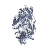
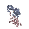
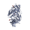
| Title | Stimulating state of a truncated Hsp70 DnaK fused with a substrate peptide | ||||||
 Components Components | Chaperone protein DnaK fused with substrate peptide | ||||||
 Keywords Keywords | CHAPERONE / molecular chaperone / Hsp70 / protein folding | ||||||
| Function / homology |  Function and homology information Function and homology information | ||||||
| Biological species |  | ||||||
| Method |  X-RAY DIFFRACTION / SYNCHROTRON / MOLECULAR REPLACEMENT / Resolution: 1.82 Å X-RAY DIFFRACTION / SYNCHROTRON / MOLECULAR REPLACEMENT / Resolution: 1.82 Å | ||||||
 Authors Authors | Wang, W. / Hendrickson, W.A. | ||||||
| Funding support |  United States, 1items United States, 1items
| ||||||
 Citation Citation | Journal: Mol.Cell / Year: 2021 Title: Conformational equilibria in allosteric control of Hsp70 chaperones. Authors: Wang, W. / Liu, Q. / Liu, Q. / Hendrickson, W.A. | ||||||
| History |
|
- Structure visualization
| Structure viewer | Molecule: MolmilJmol/JSmol |
|---|
- Downloads & links
-Download
| PDBx/mmCIF format | 7kru.cif.gz | 614.9 KB | Display | PDBx/mmCIF format |
|---|---|---|---|---|
| PDB format | pdb7kru.ent.gz | 518.5 KB | Display | PDB format |
| PDBx/mmJSON format | 7kru.json.gz | Tree view | PDBx/mmJSON format | |
| Others |  Other downloads Other downloads |
-Validation report
| Arichive directory | https://data.pdbj.org/pub/pdb/validation_reports/kr/7kruftp://data.pdbj.org/pub/pdb/validation_reports/kr/7kru | HTTPS FTP |
|---|
-Related structure data
| Related structure data |  7ko2C  7krtC  7krvC  7krwC 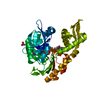 7n46C 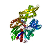 7raxC 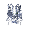 1dkzS 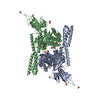 4jneS C: citing same article ( S: Starting model for refinement |
|---|---|
| Similar structure data |


-Links
 PDBj
PDBj
- Assembly
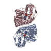
| Deposited unit | 
| |||||||||||||||||||||||||||||||||||||||||||||||||||||||||||||||||||||||||
|---|---|---|---|---|---|---|---|---|---|---|---|---|---|---|---|---|---|---|---|---|---|---|---|---|---|---|---|---|---|---|---|---|---|---|---|---|---|---|---|---|---|---|---|---|---|---|---|---|---|---|---|---|---|---|---|---|---|---|---|---|---|---|---|---|---|---|---|---|---|---|---|---|---|---|
| 1 | 
| |||||||||||||||||||||||||||||||||||||||||||||||||||||||||||||||||||||||||
| 2 | 
| |||||||||||||||||||||||||||||||||||||||||||||||||||||||||||||||||||||||||
| Unit cell |
| |||||||||||||||||||||||||||||||||||||||||||||||||||||||||||||||||||||||||
| Components on special symmetry positions |
| |||||||||||||||||||||||||||||||||||||||||||||||||||||||||||||||||||||||||
| Noncrystallographic symmetry (NCS) | NCS domain:
NCS domain segments:
|
