 Movie
Movie Controller


+ Open data

- Basic information
| Entry | Database: PDB / ID: 7n46 | ||||||
|---|---|---|---|---|---|---|---|
| Title | ADP-binding state of the nucleotide-binding domain of Hsp70 DnaK | ||||||
 Components Components | Chaperone protein DnaK | ||||||
 Keywords Keywords | CHAPERONE / molecular chaperone / Hsp70 / protein folding | ||||||
| Function / homology |  Function and homology information Function and homology information | ||||||
| Biological species |  | ||||||
| Method |  X-RAY DIFFRACTION / SYNCHROTRON / MOLECULAR REPLACEMENT / Resolution: 2.21 Å X-RAY DIFFRACTION / SYNCHROTRON / MOLECULAR REPLACEMENT / Resolution: 2.21 Å | ||||||
 Authors Authors | Wang, W. / Hendrickson, W.A. | ||||||
| Funding support |  United States, 1items United States, 1items
| ||||||
 Citation Citation | Journal: Mol.Cell / Year: 2021 Title: Conformational equilibria in allosteric control of Hsp70 chaperones. Authors: Wang, W. / Liu, Q. / Liu, Q. / Hendrickson, W.A. | ||||||
| History |
|
- Structure visualization
| Structure viewer | Molecule: MolmilJmol/JSmol |
|---|
- Downloads & links
-Download
| PDBx/mmCIF format | 7n46.cif.gz | 163.7 KB | Display | PDBx/mmCIF format |
|---|---|---|---|---|
| PDB format | pdb7n46.ent.gz | 127.5 KB | Display | PDB format |
| PDBx/mmJSON format | 7n46.json.gz | Tree view | PDBx/mmJSON format | |
| Others |  Other downloads Other downloads |
-Validation report
| Arichive directory | https://data.pdbj.org/pub/pdb/validation_reports/n4/7n46ftp://data.pdbj.org/pub/pdb/validation_reports/n4/7n46 | HTTPS FTP |
|---|
-Related structure data
| Related structure data |  7ko2C 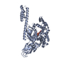 7krtC 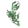 7kruC 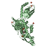 7krvC 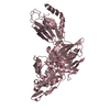 7krwC 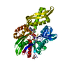 7raxC 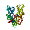 1bupS S: Starting model for refinement C: citing same article ( |
|---|---|
| Similar structure data |
-Links
 PDBj
PDBj
- Assembly
| Deposited unit | 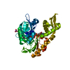
| ||||||||
|---|---|---|---|---|---|---|---|---|---|
| 1 |
| ||||||||
| Unit cell |
|
-Components
-Protein , 1 types, 1 molecules A
| #1: Protein | Mass: 41783.207 Da / Num. of mol.: 1 Source method: isolated from a genetically manipulated source Source: (gene. exp.) Strain: K12 / Gene: dnaK, FAZ83_07380 Production host: References: UniProt: A0A6D2W465 |
|---|
-Non-polymers , 5 types, 200 molecules 


| #2: Chemical | ChemComp-ADP /  Mass: 427.201 Da / Num. of mol.: 1 / Source method: obtained synthetically / Formula: C10H15N5O10P2 / Feature type: SUBJECT OF INVESTIGATION / Comment: ADP, energy-carrying molecule*YM Mass: 427.201 Da / Num. of mol.: 1 / Source method: obtained synthetically / Formula: C10H15N5O10P2 / Feature type: SUBJECT OF INVESTIGATION / Comment: ADP, energy-carrying molecule*YM |
|---|---|
| #3: Chemical | ChemComp-NO3 /  Mass: 62.005 Da / Num. of mol.: 1 / Source method: obtained synthetically / Formula: NO3 Mass: 62.005 Da / Num. of mol.: 1 / Source method: obtained synthetically / Formula: NO3 |
| #4: Chemical | ChemComp-MG /  Mass: 24.305 Da / Num. of mol.: 1 / Source method: obtained synthetically / Formula: Mg Mass: 24.305 Da / Num. of mol.: 1 / Source method: obtained synthetically / Formula: Mg |
| #5: Chemical | ChemComp-PO4 /  Mass: 94.971 Da / Num. of mol.: 1 / Source method: obtained synthetically / Formula: PO4 / Feature type: SUBJECT OF INVESTIGATION Mass: 94.971 Da / Num. of mol.: 1 / Source method: obtained synthetically / Formula: PO4 / Feature type: SUBJECT OF INVESTIGATION |
| #6: Water | ChemComp-HOH / Mass: 18.015 Da / Num. of mol.: 196 / Source method: isolated from a natural source / Formula: H2O |
-Details
| Has ligand of interest | Y |
|---|
-Experimental details
-Experiment
| Experiment | Method: X-RAY DIFFRACTION / Number of used crystals: 1 |
|---|
- Sample preparation
| Crystal | Density Matthews: 2.74 Å3/Da / Density % sol: 55.08 % |
|---|---|
| Crystal grow | Temperature: 277.15 K / Method: evaporation Details: 0.04 M HEPES pH 7.5, 20% PEG 3350, 0.1 M magnesium nitrate, 0.02 M sodium chloride, 0.01 M phosphate-citrate pH 4.2, 1% PEG 3000 |
-Data collection
| Diffraction | Mean temperature: 100 K / Serial crystal experiment: N | ||||||||||||||||||||||||||||||
|---|---|---|---|---|---|---|---|---|---|---|---|---|---|---|---|---|---|---|---|---|---|---|---|---|---|---|---|---|---|---|---|
| Diffraction source | Source: SYNCHROTRON / Site: APS / Beamline: 24-ID-C / Wavelength: 0.97918 Å | ||||||||||||||||||||||||||||||
| Detector | Type: DECTRIS PILATUS 6M / Detector: PIXEL / Date: Apr 23, 2017 | ||||||||||||||||||||||||||||||
| Radiation | Protocol: SINGLE WAVELENGTH / Monochromatic (M) / Laue (L): M / Scattering type: x-ray | ||||||||||||||||||||||||||||||
| Radiation wavelength | Wavelength: 0.97918 Å / Relative weight: 1 | ||||||||||||||||||||||||||||||
| Reflection | Resolution: 2.21→48.25 Å / Num. obs: 24196 / % possible obs: 99.9 % / Redundancy: 10.6 % / CC1/2: 0.96 / Rmerge(I) obs: 0.625 / Rpim(I) all: 0.199 / Rrim(I) all: 0.657 / Net I/σ(I): 3.7 / Num. measured all: 256415 | ||||||||||||||||||||||||||||||
| Reflection shell | Diffraction-ID: 1
|
- Processing
| Software |
| ||||||||||||||||||||||||||||||||||||||||||||||||||||||||||||||||||||||||||||||||||||||||||||||||||||
|---|---|---|---|---|---|---|---|---|---|---|---|---|---|---|---|---|---|---|---|---|---|---|---|---|---|---|---|---|---|---|---|---|---|---|---|---|---|---|---|---|---|---|---|---|---|---|---|---|---|---|---|---|---|---|---|---|---|---|---|---|---|---|---|---|---|---|---|---|---|---|---|---|---|---|---|---|---|---|---|---|---|---|---|---|---|---|---|---|---|---|---|---|---|---|---|---|---|---|---|---|---|
| Refinement | Method to determine structure: MOLECULAR REPLACEMENT Starting model: 1BUP Resolution: 2.21→48.25 Å / SU ML: 0.28 / Cross valid method: THROUGHOUT / σ(F): 1.34 / Phase error: 24.04 / Stereochemistry target values: ML
| ||||||||||||||||||||||||||||||||||||||||||||||||||||||||||||||||||||||||||||||||||||||||||||||||||||
| Solvent computation | Shrinkage radii: 0.9 Å / VDW probe radii: 1.11 Å / Solvent model: FLAT BULK SOLVENT MODEL | ||||||||||||||||||||||||||||||||||||||||||||||||||||||||||||||||||||||||||||||||||||||||||||||||||||
| Displacement parameters | Biso max: 107.97 Å2 / Biso mean: 30.1686 Å2 / Biso min: 13.02 Å2 | ||||||||||||||||||||||||||||||||||||||||||||||||||||||||||||||||||||||||||||||||||||||||||||||||||||
| Refinement step | Cycle: final / Resolution: 2.21→48.25 Å
| ||||||||||||||||||||||||||||||||||||||||||||||||||||||||||||||||||||||||||||||||||||||||||||||||||||
| Refine LS restraints |
| ||||||||||||||||||||||||||||||||||||||||||||||||||||||||||||||||||||||||||||||||||||||||||||||||||||
| LS refinement shell | Refine-ID: X-RAY DIFFRACTION / Rfactor Rfree error: 0 / Total num. of bins used: 8
| ||||||||||||||||||||||||||||||||||||||||||||||||||||||||||||||||||||||||||||||||||||||||||||||||||||
| Refinement TLS params. | Method: refined / Refine-ID: X-RAY DIFFRACTION
| ||||||||||||||||||||||||||||||||||||||||||||||||||||||||||||||||||||||||||||||||||||||||||||||||||||
| Refinement TLS group |
|
