 Movie
Movie Controller

[English] 日本語
 Yorodumi
- PDB-7kpj: Crystal structure of Ruminococcus gnavus immunoglobulin binding p... -
+ Open data

- Basic information
| Entry | Database: PDB / ID: 7kpj | |||||||||
|---|---|---|---|---|---|---|---|---|---|---|
| Title | Crystal structure of Ruminococcus gnavus immunoglobulin binding protein in complex with 338E6 Fab | |||||||||
 Components Components |
| |||||||||
 Keywords Keywords | IMMUNE SYSTEM / superantigen / immunoglobulin binding protein / immunoglobulin complex | |||||||||
| Function / homology | cell wall / Gram-positive cocci surface proteins LPxTG motif profile. / LPXTG cell wall anchor domain / membrane => GO:0016020 / extracellular region / GRAM_POS_ANCHORING domain-containing protein Function and homology information Function and homology information | |||||||||
| Biological species |   Ruminococcus gnavus (bacteria) Ruminococcus gnavus (bacteria) | |||||||||
| Method |  X-RAY DIFFRACTION / SYNCHROTRON / MOLECULAR REPLACEMENT / Resolution: 2.1 Å X-RAY DIFFRACTION / SYNCHROTRON / MOLECULAR REPLACEMENT / Resolution: 2.1 Å | |||||||||
 Authors Authors | Borowska, M.T. / Adams, E.J. | |||||||||
| Funding support |  United States, 2items United States, 2items
| |||||||||
 Citation Citation | Journal: Proc.Natl.Acad.Sci.USA / Year: 2021 Title: The molecular characterization of antibody binding to a superantigen-like protein from a commensal microbe. Authors: Borowska, M.T. / Drees, C. / Yarawsky, A.E. / Viswanathan, M. / Ryan, S.M. / Bunker, J.J. / Herr, A.B. / Bendelac, A. / Adams, E.J. | |||||||||
| History |
|
- Structure visualization
| Structure viewer | Molecule: MolmilJmol/JSmol |
|---|
- Downloads & links
-Download
| PDBx/mmCIF format | 7kpj.cif.gz | 500.2 KB | Display | PDBx/mmCIF format |
|---|---|---|---|---|
| PDB format | pdb7kpj.ent.gz | 408 KB | Display | PDB format |
| PDBx/mmJSON format | 7kpj.json.gz | Tree view | PDBx/mmJSON format | |
| Others |  Other downloads Other downloads |
-Validation report
| Summary document | 7kpj_validation.pdf.gz | 474.1 KB | Display | wwPDB validaton report |
|---|---|---|---|---|
| Full document | 7kpj_full_validation.pdf.gz | 493 KB | Display | |
| Data in XML | 7kpj_validation.xml.gz | 46.6 KB | Display | |
| Data in CIF | 7kpj_validation.cif.gz | 66 KB | Display | |
| Arichive directory | https://data.pdbj.org/pub/pdb/validation_reports/kp/7kpjftp://data.pdbj.org/pub/pdb/validation_reports/kp/7kpj | HTTPS FTP |
-Related structure data
| Related structure data |  1deeS S: Starting model for refinement |
|---|---|
| Similar structure data |


-Links
 PDBj
PDBj

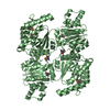
- Assembly
| Deposited unit | 
| ||||||||
|---|---|---|---|---|---|---|---|---|---|
| 1 | 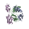
| ||||||||
| 2 | 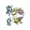
| ||||||||
| Unit cell |
|
-Components
| #1: Antibody | Mass: 23472.873 Da / Num. of mol.: 2 Source method: isolated from a genetically manipulated source Source: (gene. exp.)  Trichoplusia ni (cabbage looper) Trichoplusia ni (cabbage looper)#2: Protein | Mass: 30495.400 Da / Num. of mol.: 2 Source method: isolated from a genetically manipulated source Source: (gene. exp.) Ruminococcus gnavus (bacteria) / Gene: CDL27_16280 / Production host: #3: Antibody | Mass: 24062.938 Da / Num. of mol.: 2 Source method: isolated from a genetically manipulated source Source: (gene. exp.) Trichoplusia ni (cabbage looper)#4: Chemical |   Mass: 40.078 Da / Num. of mol.: 2 / Source method: obtained synthetically / Formula: Ca Mass: 40.078 Da / Num. of mol.: 2 / Source method: obtained synthetically / Formula: Ca#5: Water | ChemComp-HOH / |  Mass: 18.015 Da / Num. of mol.: 273 / Source method: isolated from a natural source / Formula: H2O Mass: 18.015 Da / Num. of mol.: 273 / Source method: isolated from a natural source / Formula: H2OHas ligand of interest | N | |
|---|
-Experimental details
-Experiment
| Experiment | Method: X-RAY DIFFRACTION / Number of used crystals: 1 |
|---|
- Sample preparation
| Crystal | Density Matthews: 3.23 Å3/Da / Density % sol: 61.9 % |
|---|---|
| Crystal grow | Temperature: 298 K / Method: vapor diffusion, hanging drop / pH: 7.4 / Details: 0.2 M calcium chloride, 18% PEG 4000 |
-Data collection
| Diffraction | Mean temperature: 100 K / Serial crystal experiment: N |
|---|---|
| Diffraction source | Source: SYNCHROTRON / Site: APS / Beamline: 24-ID-E / Wavelength: 0.9791 Å |
| Detector | Type: DECTRIS EIGER X 16M / Detector: PIXEL / Date: Dec 9, 2018 |
| Radiation | Protocol: SINGLE WAVELENGTH / Monochromatic (M) / Laue (L): M / Scattering type: x-ray |
| Radiation wavelength | Wavelength: 0.9791 Å / Relative weight: 1 |
| Reflection | Resolution: 2.1→83.36 Å / Num. obs: 115284 / % possible obs: 99.1 % / Redundancy: 33.4 % / CC1/2: 0.971 / CC star: 0.993 / Net I/σ(I): 21.03 |
| Reflection shell | Resolution: 2.1→2.177 Å / Redundancy: 32.5 % / Num. unique obs: 11391 / CC1/2: 0.307 / CC star: 0.686 / % possible all: 98.16 |
- Processing
| Software |
| |||||||||||||||||||||||||||||||||||||||||||||||||||||||||||||||||||||||||||||||||||||||||||||||||||||||||
|---|---|---|---|---|---|---|---|---|---|---|---|---|---|---|---|---|---|---|---|---|---|---|---|---|---|---|---|---|---|---|---|---|---|---|---|---|---|---|---|---|---|---|---|---|---|---|---|---|---|---|---|---|---|---|---|---|---|---|---|---|---|---|---|---|---|---|---|---|---|---|---|---|---|---|---|---|---|---|---|---|---|---|---|---|---|---|---|---|---|---|---|---|---|---|---|---|---|---|---|---|---|---|---|---|---|---|
| Refinement | Method to determine structure: MOLECULAR REPLACEMENT Starting model: 1dee Resolution: 2.1→83.36 Å / SU ML: 0.35 / Cross valid method: THROUGHOUT / σ(F): 1.34 / Phase error: 31.11 / Stereochemistry target values: ML
| |||||||||||||||||||||||||||||||||||||||||||||||||||||||||||||||||||||||||||||||||||||||||||||||||||||||||
| Solvent computation | Shrinkage radii: 0.9 Å / VDW probe radii: 1.11 Å / Solvent model: FLAT BULK SOLVENT MODEL | |||||||||||||||||||||||||||||||||||||||||||||||||||||||||||||||||||||||||||||||||||||||||||||||||||||||||
| Displacement parameters | Biso max: 116.59 Å2 / Biso mean: 48.7058 Å2 / Biso min: 19.89 Å2 | |||||||||||||||||||||||||||||||||||||||||||||||||||||||||||||||||||||||||||||||||||||||||||||||||||||||||
| Refinement step | Cycle: final / Resolution: 2.1→83.36 Å
| |||||||||||||||||||||||||||||||||||||||||||||||||||||||||||||||||||||||||||||||||||||||||||||||||||||||||
| Refine LS restraints |
| |||||||||||||||||||||||||||||||||||||||||||||||||||||||||||||||||||||||||||||||||||||||||||||||||||||||||
| LS refinement shell | Refine-ID: X-RAY DIFFRACTION / Rfactor Rfree error: 0 / Total num. of bins used: 14
| |||||||||||||||||||||||||||||||||||||||||||||||||||||||||||||||||||||||||||||||||||||||||||||||||||||||||
| Refinement TLS params. | Method: refined / Origin x: -2.8509 Å / Origin y: -20.9307 Å / Origin z: -34.2326 Å
| |||||||||||||||||||||||||||||||||||||||||||||||||||||||||||||||||||||||||||||||||||||||||||||||||||||||||
| Refinement TLS group |
|
