 Movie
Movie Controller


+ Open data

- Basic information
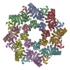
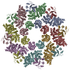
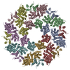
| Entry | Database: PDB / ID: 7knq | |||||||||||||||||||||||||||
|---|---|---|---|---|---|---|---|---|---|---|---|---|---|---|---|---|---|---|---|---|---|---|---|---|---|---|---|---|
| Title | SARM1 Octamer | |||||||||||||||||||||||||||
 Components Components | NAD(+) hydrolase SARM1 | |||||||||||||||||||||||||||
 Keywords Keywords | HYDROLASE / homo-oligomer / Mitochondria localized protein | |||||||||||||||||||||||||||
| Function / homology |  Function and homology information Function and homology informationnegative regulation of MyD88-independent toll-like receptor signaling pathway / extrinsic component of synaptic membrane / MyD88-independent TLR4 cascade / NADP+ nucleosidase activity / Toll Like Receptor 3 (TLR3) Cascade / NAD+ nucleosidase activity / regulation of synapse pruning / modification of postsynaptic structure / NAD+ catabolic process / ADP-ribosyl cyclase/cyclic ADP-ribose hydrolase ...negative regulation of MyD88-independent toll-like receptor signaling pathway / extrinsic component of synaptic membrane / MyD88-independent TLR4 cascade / NADP+ nucleosidase activity / Toll Like Receptor 3 (TLR3) Cascade / NAD+ nucleosidase activity / regulation of synapse pruning / modification of postsynaptic structure / NAD+ catabolic process / ADP-ribosyl cyclase/cyclic ADP-ribose hydrolase / NAD+ nucleosidase activity, cyclic ADP-ribose generating / protein localization to mitochondrion / nervous system process / Hydrolases; Glycosylases; Hydrolysing N-glycosyl compounds / regulation of dendrite morphogenesis / response to axon injury / response to glucose / signaling adaptor activity / regulation of neuron apoptotic process / TRAF6-mediated induction of TAK1 complex within TLR4 complex / Activation of IRF3, IRF7 mediated by TBK1, IKKε (IKBKE) / IKK complex recruitment mediated by RIP1 / neuromuscular junction / nervous system development / microtubule / cell differentiation / mitochondrial outer membrane / innate immune response / axon / synapse / dendrite / glutamatergic synapse / cell surface / signal transduction / protein-containing complex / mitochondrion / identical protein binding / cytoplasm / cytosol Similarity search - Function | |||||||||||||||||||||||||||
| Biological species |  Homo sapiens (human) Homo sapiens (human) | |||||||||||||||||||||||||||
| Method | ELECTRON MICROSCOPY / single particle reconstruction / cryo EM / Resolution: 3.4 Å | |||||||||||||||||||||||||||
 Authors Authors | Shen, C. / Wu, H. | |||||||||||||||||||||||||||
| Funding support |  United States, 1items United States, 1items
| |||||||||||||||||||||||||||
 Citation Citation | Journal: Proc Natl Acad Sci U S A / Year: 2021 Title: Multiple domain interfaces mediate SARM1 autoinhibition. Authors: Chen Shen / Mihir Vohra / Pengfei Zhang / Xianrong Mao / Matthew D Figley / Jian Zhu / Yo Sasaki / Hao Wu / Aaron DiAntonio / Jeffrey Milbrandt / Abstract: Axon degeneration is an active program of self-destruction mediated by the protein SARM1. In healthy neurons, SARM1 is autoinhibited and, upon injury autoinhibition is relieved, activating the SARM1 ...Axon degeneration is an active program of self-destruction mediated by the protein SARM1. In healthy neurons, SARM1 is autoinhibited and, upon injury autoinhibition is relieved, activating the SARM1 enzyme to deplete NAD and induce axon degeneration. SARM1 forms a homomultimeric octamer with each monomer composed of an N-terminal autoinhibitory ARM domain, tandem SAM domains that mediate multimerization, and a C-terminal TIR domain encoding the NADase enzyme. Here we discovered multiple intramolecular and intermolecular domain interfaces required for SARM1 autoinhibition using peptide mapping and cryo-electron microscopy (cryo-EM). We identified a candidate autoinhibitory region by screening a panel of peptides derived from the SARM1 ARM domain, identifying a peptide mediating high-affinity inhibition of the SARM1 NADase. Mutation of residues in full-length SARM1 within the region encompassed by the peptide led to loss of autoinhibition, rendering SARM1 constitutively active and inducing spontaneous NAD and axon loss. The cryo-EM structure of SARM1 revealed 1) a compact autoinhibited SARM1 octamer in which the TIR domains are isolated and prevented from oligomerization and enzymatic activation and 2) multiple candidate autoinhibitory interfaces among the domains. Mutational analysis demonstrated that five distinct interfaces are required for autoinhibition, including intramolecular and intermolecular ARM-SAM interfaces, an intermolecular ARM-ARM interface, and two ARM-TIR interfaces formed between a single TIR and two distinct ARM domains. These autoinhibitory regions are not redundant, as point mutants in each led to constitutively active SARM1. These studies define the structural basis for SARM1 autoinhibition and may enable the development of SARM1 inhibitors that stabilize the autoinhibited state. | |||||||||||||||||||||||||||
| History |
|
- Structure visualization
| Movie |
Movie viewer |
|---|---|
| Structure viewer | Molecule: MolmilJmol/JSmol |
- Downloads & links
-Download
| PDBx/mmCIF format | 7knq.cif.gz | 935.2 KB | Display | PDBx/mmCIF format |
|---|---|---|---|---|
| PDB format | pdb7knq.ent.gz | 756.1 KB | Display | PDB format |
| PDBx/mmJSON format | 7knq.json.gz | Tree view | PDBx/mmJSON format | |
| Others |  Other downloads Other downloads |
-Validation report
| Arichive directory | https://data.pdbj.org/pub/pdb/validation_reports/kn/7knqftp://data.pdbj.org/pub/pdb/validation_reports/kn/7knq | HTTPS FTP |
|---|
-Related structure data
| Related structure data | 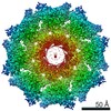 22954MC M: map data used to model this data C: citing same article ( |
|---|---|
| Similar structure data |


-Links
 PDBj
PDBj


- Assembly
| Deposited unit | 
|
|---|---|
| 1 |
|
-Components
| #1: Protein | Mass: 79486.164 Da / Num. of mol.: 8 Source method: isolated from a genetically manipulated source Source: (gene. exp.) Homo sapiens (human) / Gene: SARM1, KIAA0524, SAMD2, SARM / Production host:   Spodoptera frugiperda (fall armyworm) Spodoptera frugiperda (fall armyworm)References: UniProt: Q6SZW1, ADP-ribosyl cyclase/cyclic ADP-ribose hydrolase, Hydrolases; Glycosylases; Hydrolysing N-glycosyl compounds Has protein modification | N | |
|---|
-Experimental details
-Experiment
| Experiment | Method: ELECTRON MICROSCOPY |
|---|---|
| EM experiment | Aggregation state: PARTICLE / 3D reconstruction method: single particle reconstruction |
- Sample preparation
| Component | Name: Octameric form of SARM1 / Type: COMPLEX / Entity ID: all / Source: RECOMBINANT |
|---|---|
| Source (natural) | Organism: Homo sapiens (human) |
| Source (recombinant) | Organism: Spodoptera frugiperda (fall armyworm) |
| Buffer solution | pH: 7.4 |
| Specimen | Embedding applied: NO / Shadowing applied: NO / Staining applied: NO / Vitrification applied: YES |
| Vitrification | Cryogen name: ETHANE |
- Electron microscopy imaging
| Experimental equipment |  Model: Titan Krios / Image courtesy: FEI Company |
|---|---|
| Microscopy | Model: FEI TITAN KRIOS |
| Electron gun | Electron source:  FIELD EMISSION GUN / Accelerating voltage: 300 kV / Illumination mode: FLOOD BEAM FIELD EMISSION GUN / Accelerating voltage: 300 kV / Illumination mode: FLOOD BEAM |
| Electron lens | Mode: BRIGHT FIELD |
| Image recording | Electron dose: 69.2 e/Å2 / Film or detector model: GATAN K3 (6k x 4k) |
- Processing
| Software | Name: PHENIX / Version: 1.18.2_3874: / Classification: refinement | ||||||||||||||||||||||||
|---|---|---|---|---|---|---|---|---|---|---|---|---|---|---|---|---|---|---|---|---|---|---|---|---|---|
| EM software | Name: PHENIX / Category: model refinement | ||||||||||||||||||||||||
| CTF correction | Type: NONE | ||||||||||||||||||||||||
| 3D reconstruction | Resolution: 3.4 Å / Resolution method: FSC 0.143 CUT-OFF / Num. of particles: 34643 / Symmetry type: POINT | ||||||||||||||||||||||||
| Refine LS restraints |
|
