 Movie
Movie Controller

[English] 日本語
 Yorodumi
- PDB-7kn8: Crystal structure of the GH74 xyloglucanase from Xanthomonas camp... -
+ Open data

- Basic information
| Entry | Database: PDB / ID: 7kn8 | |||||||||||||||
|---|---|---|---|---|---|---|---|---|---|---|---|---|---|---|---|---|
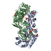
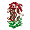
| Title | Crystal structure of the GH74 xyloglucanase from Xanthomonas campestris (Xcc1752) | |||||||||||||||
 Components Components | Cellulase | |||||||||||||||
 Keywords Keywords | HYDROLASE / Glycoside Hydrolase Family 74 / Xyloglucanase / Xyloglucan | |||||||||||||||
| Function / homology | : / xyloglucan metabolic process / hydrolase activity, acting on glycosyl bonds / polysaccharide catabolic process / WD40/YVTN repeat-like-containing domain superfamily / IODIDE ION / Cellulase Function and homology information Function and homology information | |||||||||||||||
| Biological species |  Xanthomonas campestris pv. campestris (bacteria) Xanthomonas campestris pv. campestris (bacteria) | |||||||||||||||
| Method |  X-RAY DIFFRACTION / SYNCHROTRON / MOLECULAR REPLACEMENT / Resolution: 1.95 Å X-RAY DIFFRACTION / SYNCHROTRON / MOLECULAR REPLACEMENT / Resolution: 1.95 Å | |||||||||||||||
 Authors Authors | Araujo, E.A. / Vieira, P.S. / Murakami, M.T. / Polikarpov, I. | |||||||||||||||
| Funding support |  Brazil, 4items Brazil, 4items
| |||||||||||||||
 Citation Citation | Journal: Nature Communications / Year: 2021 Title: Xyloglucan processing machinery in Xanthomonas pathogens and its role in the transcriptional activation of virulence factors Authors: Vieira, P.S. / Bonfim, I.M. / Araujo, E.A. / Melo, R.R. / Lima, A.R. / Fessel, M.R. / Paixao, D.A.A. / Persinoti, G.F. / Rocco, S.A. / Lima, T.B. / Pirolla, R.A.S. / Morais, M.A.B. / Correa, ...Authors: Vieira, P.S. / Bonfim, I.M. / Araujo, E.A. / Melo, R.R. / Lima, A.R. / Fessel, M.R. / Paixao, D.A.A. / Persinoti, G.F. / Rocco, S.A. / Lima, T.B. / Pirolla, R.A.S. / Morais, M.A.B. / Correa, J.B.L. / Zanphorlin, L.M. / Diogo, J.A. / Lima, E.A. / Grandis, A. / Buckeridge, M.S. / Gozzo, F.C. / Benedetti, C.E. / Polikarpov, I. / Giuseppe, P.O. / Murakami, M.T. | |||||||||||||||
| History |
|
- Structure visualization
| Structure viewer | Molecule: MolmilJmol/JSmol |
|---|
- Downloads & links
-Download
| PDBx/mmCIF format | 7kn8.cif.gz | 541 KB | Display | PDBx/mmCIF format |
|---|---|---|---|---|
| PDB format | pdb7kn8.ent.gz | 440.3 KB | Display | PDB format |
| PDBx/mmJSON format | 7kn8.json.gz | Tree view | PDBx/mmJSON format | |
| Others |  Other downloads Other downloads |
-Validation report
| Arichive directory | https://data.pdbj.org/pub/pdb/validation_reports/kn/7kn8ftp://data.pdbj.org/pub/pdb/validation_reports/kn/7kn8 | HTTPS FTP |
|---|
-Related structure data
| Related structure data |  7kmmC  7kmnC  7kmoC 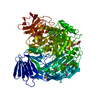 7kmpC  7kmqC 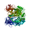 7kncC 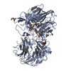 2cn2S S: Starting model for refinement C: citing same article ( |
|---|---|
| Similar structure data |


-Links
 PDBj
PDBj- Assembly
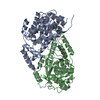
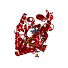
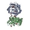
| Deposited unit | 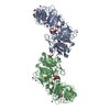
| ||||||||
|---|---|---|---|---|---|---|---|---|---|
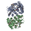
| 1 | 
| ||||||||
| 2 | 
| ||||||||
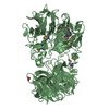
| Unit cell |
|
-Components
-Protein / Sugars , 2 types, 4 molecules AB
| #1: Protein | Mass: 77306.688 Da / Num. of mol.: 2 Source method: isolated from a genetically manipulated source Source: (gene. exp.) Xanthomonas campestris pv. campestris (strain ATCC 33913 / DSM 3586 / NCPPB 528 / LMG 568 / P 25) (bacteria)Strain: ATCC 33913 / DSM 3586 / NCPPB 528 / LMG 568 / P 25 / Gene: XCC1752 / Production host: #2: Polysaccharide | Source method: isolated from a genetically manipulated source |
|---|
-Non-polymers , 4 types, 1109 molecules 


| #3: Chemical | ChemComp-EDO /  Mass: 62.068 Da / Num. of mol.: 9 / Source method: obtained synthetically / Formula: C2H6O2 Mass: 62.068 Da / Num. of mol.: 9 / Source method: obtained synthetically / Formula: C2H6O2#4: Chemical |  Mass: 22.990 Da / Num. of mol.: 2 / Source method: obtained synthetically / Formula: Na Mass: 22.990 Da / Num. of mol.: 2 / Source method: obtained synthetically / Formula: Na#5: Chemical | ChemComp-IOD /  Mass: 126.904 Da / Num. of mol.: 14 / Source method: obtained synthetically / Formula: I Mass: 126.904 Da / Num. of mol.: 14 / Source method: obtained synthetically / Formula: I#6: Water | ChemComp-HOH / | Mass: 18.015 Da / Num. of mol.: 1084 / Source method: isolated from a natural source / Formula: H2O |
|---|
-Details
| Has ligand of interest | Y |
|---|
-Experimental details
-Experiment
| Experiment | Method: X-RAY DIFFRACTION / Number of used crystals: 1 |
|---|
- Sample preparation
| Crystal | Density Matthews: 2.23 Å3/Da / Density % sol: 44.97 % |
|---|---|
| Crystal grow | Temperature: 291 K / Method: vapor diffusion, sitting drop / pH: 7.5 Details: 0.2 mol/L sodium iodede 0.1 mol/L bis-tris propane 20% PEG 3350 |
-Data collection
| Diffraction | Mean temperature: 100 K / Serial crystal experiment: N |
|---|---|
| Diffraction source | Source: SYNCHROTRON / Site: LNLS / Beamline: W01B-MX2 / Wavelength: 1.45855 Å |
| Detector | Type: DECTRIS PILATUS 2M / Detector: PIXEL / Date: Oct 23, 2012 |
| Radiation | Monochromator: Water-cooled Si(111) / Protocol: SINGLE WAVELENGTH / Monochromatic (M) / Laue (L): M / Scattering type: x-ray |
| Radiation wavelength | Wavelength: 1.45855 Å / Relative weight: 1 |
| Reflection | Resolution: 1.95→19.992 Å / Num. obs: 99662 / % possible obs: 98.1 % / Redundancy: 12.191 % / CC1/2: 0.997 / Net I/σ(I): 11.02 |
| Reflection shell | Resolution: 1.95→2.07 Å / Redundancy: 10.12 % / Mean I/σ(I) obs: 2.5 / Num. unique obs: 15178 / CC1/2: 0.888 / % possible all: 93.7 |
- Processing
| Software |
| ||||||||||||||||||||||||||||||||||||||||||||||||||||||||||||||||||||||||||||||||||||||||||||||||||||||||||||||||||||||||||||||||||||||||||||||||||||||||||||||||||||||||||||||||||||||||||
|---|---|---|---|---|---|---|---|---|---|---|---|---|---|---|---|---|---|---|---|---|---|---|---|---|---|---|---|---|---|---|---|---|---|---|---|---|---|---|---|---|---|---|---|---|---|---|---|---|---|---|---|---|---|---|---|---|---|---|---|---|---|---|---|---|---|---|---|---|---|---|---|---|---|---|---|---|---|---|---|---|---|---|---|---|---|---|---|---|---|---|---|---|---|---|---|---|---|---|---|---|---|---|---|---|---|---|---|---|---|---|---|---|---|---|---|---|---|---|---|---|---|---|---|---|---|---|---|---|---|---|---|---|---|---|---|---|---|---|---|---|---|---|---|---|---|---|---|---|---|---|---|---|---|---|---|---|---|---|---|---|---|---|---|---|---|---|---|---|---|---|---|---|---|---|---|---|---|---|---|---|---|---|---|---|---|---|---|
| Refinement | Method to determine structure: MOLECULAR REPLACEMENT Starting model: 2CN2 Resolution: 1.95→19.992 Å / SU ML: 0.25 / Cross valid method: THROUGHOUT / σ(F): 1.35 / Phase error: 29.53 / Stereochemistry target values: ML
| ||||||||||||||||||||||||||||||||||||||||||||||||||||||||||||||||||||||||||||||||||||||||||||||||||||||||||||||||||||||||||||||||||||||||||||||||||||||||||||||||||||||||||||||||||||||||||
| Solvent computation | Shrinkage radii: 0.9 Å / VDW probe radii: 1.11 Å / Solvent model: FLAT BULK SOLVENT MODEL | ||||||||||||||||||||||||||||||||||||||||||||||||||||||||||||||||||||||||||||||||||||||||||||||||||||||||||||||||||||||||||||||||||||||||||||||||||||||||||||||||||||||||||||||||||||||||||
| Displacement parameters | Biso max: 64.54 Å2 / Biso mean: 19.4244 Å2 / Biso min: 14.26 Å2 | ||||||||||||||||||||||||||||||||||||||||||||||||||||||||||||||||||||||||||||||||||||||||||||||||||||||||||||||||||||||||||||||||||||||||||||||||||||||||||||||||||||||||||||||||||||||||||
| Refinement step | Cycle: final / Resolution: 1.95→19.992 Å
| ||||||||||||||||||||||||||||||||||||||||||||||||||||||||||||||||||||||||||||||||||||||||||||||||||||||||||||||||||||||||||||||||||||||||||||||||||||||||||||||||||||||||||||||||||||||||||
| LS refinement shell | Refine-ID: X-RAY DIFFRACTION / Rfactor Rfree error: 0
|
