 Movie
Movie Controller

[English] 日本語
 Yorodumi
- PDB-7kn5: Crystal structure of SARS-CoV-2 receptor binding domain complexed... -
+ Open data

- Basic information
| Entry | Database: PDB / ID: 7kn5 | |||||||||
|---|---|---|---|---|---|---|---|---|---|---|
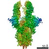
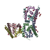
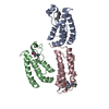
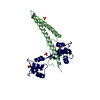
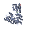
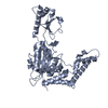
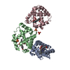
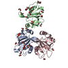
| Title | Crystal structure of SARS-CoV-2 receptor binding domain complexed with nanobodies VHH E and U | |||||||||
 Components Components |
| |||||||||
 Keywords Keywords | VIRAL PROTEIN/IMMUNE SYSTEM / SARS-CoV-2 / Nanobody / Spike / Coronavirus / COVID-19 / IMMUNE SYSTEM / Nanobody-antigen complex / single-domain antibody / VIRAL PROTEIN / VIRAL PROTEIN-IMMUNE SYSTEM complex | |||||||||
| Function / homology |  Function and homology information Function and homology informationsymbiont-mediated disruption of host tissue / Maturation of spike protein / Translation of Structural Proteins / Virion Assembly and Release / host cell surface / host extracellular region / symbiont-mediated-mediated suppression of host tetherin activity / Induction of Cell-Cell Fusion / structural constituent of virion / positive regulation of viral entry into host cell ...symbiont-mediated disruption of host tissue / Maturation of spike protein / Translation of Structural Proteins / Virion Assembly and Release / host cell surface / host extracellular region / symbiont-mediated-mediated suppression of host tetherin activity / Induction of Cell-Cell Fusion / structural constituent of virion / positive regulation of viral entry into host cell / membrane fusion / host cell endoplasmic reticulum-Golgi intermediate compartment membrane / Attachment and Entry / entry receptor-mediated virion attachment to host cell / receptor-mediated virion attachment to host cell / host cell surface receptor binding / symbiont-mediated suppression of host innate immune response / endocytosis involved in viral entry into host cell / receptor ligand activity / fusion of virus membrane with host plasma membrane / fusion of virus membrane with host endosome membrane / viral envelope / symbiont entry into host cell / virion attachment to host cell / host cell plasma membrane / SARS-CoV-2 activates/modulates innate and adaptive immune responses / virion membrane / membrane / identical protein binding / plasma membrane Similarity search - Function | |||||||||
| Biological species |   Severe acute respiratory syndrome coronavirus 2 Severe acute respiratory syndrome coronavirus 2 | |||||||||
| Method |  X-RAY DIFFRACTION / SYNCHROTRON / MOLECULAR REPLACEMENT / Resolution: 1.87 Å X-RAY DIFFRACTION / SYNCHROTRON / MOLECULAR REPLACEMENT / Resolution: 1.87 Å | |||||||||
 Authors Authors | Liu, H. / Yuan, M. / Zhu, X. / Wu, N.C. / Wilson, I.A. | |||||||||
| Funding support |  United States, 2items United States, 2items
| |||||||||
 Citation Citation | Journal: Science / Year: 2021 Title: Structure-guided multivalent nanobodies block SARS-CoV-2 infection and suppress mutational escape. Authors: Paul-Albert Koenig / Hrishikesh Das / Hejun Liu / Beate M Kümmerer / Florian N Gohr / Lea-Marie Jenster / Lisa D J Schiffelers / Yonas M Tesfamariam / Miki Uchima / Jennifer D Wuerth / Karl ...Authors: Paul-Albert Koenig / Hrishikesh Das / Hejun Liu / Beate M Kümmerer / Florian N Gohr / Lea-Marie Jenster / Lisa D J Schiffelers / Yonas M Tesfamariam / Miki Uchima / Jennifer D Wuerth / Karl Gatterdam / Natalia Ruetalo / Maria H Christensen / Caroline I Fandrey / Sabine Normann / Jan M P Tödtmann / Steffen Pritzl / Leo Hanke / Jannik Boos / Meng Yuan / Xueyong Zhu / Jonathan L Schmid-Burgk / Hiroki Kato / Michael Schindler / Ian A Wilson / Matthias Geyer / Kerstin U Ludwig / B Martin Hällberg / Nicholas C Wu / Florian I Schmidt /   Abstract: The pandemic caused by severe acute respiratory syndrome coronavirus 2 (SARS-CoV-2) continues to spread, with devastating consequences. For passive immunization efforts, nanobodies have size and cost ...The pandemic caused by severe acute respiratory syndrome coronavirus 2 (SARS-CoV-2) continues to spread, with devastating consequences. For passive immunization efforts, nanobodies have size and cost advantages over conventional antibodies. In this study, we generated four neutralizing nanobodies that target the receptor binding domain of the SARS-CoV-2 spike protein. We used x-ray crystallography and cryo-electron microscopy to define two distinct binding epitopes. On the basis of these structures, we engineered multivalent nanobodies with more than 100 times the neutralizing activity of monovalent nanobodies. Biparatopic nanobody fusions suppressed the emergence of escape mutants. Several nanobody constructs neutralized through receptor binding competition, whereas other monovalent and biparatopic nanobodies triggered aberrant activation of the spike fusion machinery. These premature conformational changes in the spike protein forestalled productive fusion and rendered the virions noninfectious. | |||||||||
| History |
|
- Structure visualization
| Structure viewer | Molecule: MolmilJmol/JSmol |
|---|
- Downloads & links
-Download
| PDBx/mmCIF format | 7kn5.cif.gz | 437 KB | Display | PDBx/mmCIF format |
|---|---|---|---|---|
| PDB format | pdb7kn5.ent.gz | 297.5 KB | Display | PDB format |
| PDBx/mmJSON format | 7kn5.json.gz | Tree view | PDBx/mmJSON format | |
| Others |  Other downloads Other downloads |
-Validation report
| Arichive directory | https://data.pdbj.org/pub/pdb/validation_reports/kn/7kn5ftp://data.pdbj.org/pub/pdb/validation_reports/kn/7kn5 | HTTPS FTP |
|---|
-Related structure data
| Related structure data | 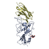 7b14C  7b17C  7b18C 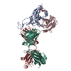 7kn6C 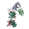 7kn7C  7ksgC 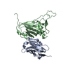 6waqS 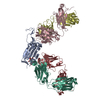 6xc7S S: Starting model for refinement C: citing same article ( |
|---|---|
| Similar structure data |


-Links
 PDBj
PDBj


- Assembly
| Deposited unit | 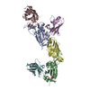
| ||||||||||||||||||||||||||||||||||||||||||||||||||||||||||||||||||||||||||||||
|---|---|---|---|---|---|---|---|---|---|---|---|---|---|---|---|---|---|---|---|---|---|---|---|---|---|---|---|---|---|---|---|---|---|---|---|---|---|---|---|---|---|---|---|---|---|---|---|---|---|---|---|---|---|---|---|---|---|---|---|---|---|---|---|---|---|---|---|---|---|---|---|---|---|---|---|---|---|---|---|
| 1 | 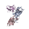
| ||||||||||||||||||||||||||||||||||||||||||||||||||||||||||||||||||||||||||||||
| 2 | 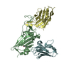
| ||||||||||||||||||||||||||||||||||||||||||||||||||||||||||||||||||||||||||||||
| Unit cell |
| ||||||||||||||||||||||||||||||||||||||||||||||||||||||||||||||||||||||||||||||
| Noncrystallographic symmetry (NCS) | NCS domain:
NCS domain segments:
|