 Movie
Movie Controller

[English] 日本語
 Yorodumi
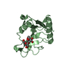
- PDB-7kij: Muscovy duck circovirus Rep domain complexed with a single-strand... -
+ Open data

- Basic information
| Entry | Database: PDB / ID: 7kij | |||||||||
|---|---|---|---|---|---|---|---|---|---|---|
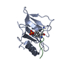
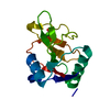
| Title | Muscovy duck circovirus Rep domain complexed with a single-stranded DNA 10-mer comprising the cleavage site | |||||||||
 Components Components |
| |||||||||
 Keywords Keywords | REPLICATION/DNA / HUH-tag / HUH motif / Rep domain / viral protein / single stranded DNA / ssDNA / ssDNA binding / REPLICATION / DNA BINDING PROTEIN / REPLICATION-DNA complex | |||||||||
| Function / homology |  Function and homology information Function and homology informationnucleotidyltransferase activity / endonuclease activity / DNA replication / RNA helicase activity / hydrolase activity / host cell nucleus / DNA binding / RNA binding / ATP binding / metal ion binding Similarity search - Function | |||||||||
| Biological species |  Muscovy duck circovirus Muscovy duck circovirus | |||||||||
| Method |  X-RAY DIFFRACTION / SYNCHROTRON / MOLECULAR REPLACEMENT / Resolution: 1.69 Å X-RAY DIFFRACTION / SYNCHROTRON / MOLECULAR REPLACEMENT / Resolution: 1.69 Å | |||||||||
 Authors Authors | Tompkins, K.J. / Gordon, W.R. / Shi, K. | |||||||||
| Funding support |  United States, 2items United States, 2items
| |||||||||
 Citation Citation | Journal: Mbio / Year: 2023 Title: Watson-Crick Base-Pairing Requirements for ssDNA Recognition and Processing in Replication-Initiating HUH Endonucleases. Authors: Smiley, A.T. / Tompkins, K.J. / Pawlak, M.R. / Krueger, A.J. / Evans 3rd, R.L. / Shi, K. / Aihara, H. / Gordon, W.R. | |||||||||
| History |
|
- Structure visualization
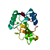
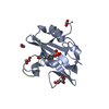
| Structure viewer | Molecule: MolmilJmol/JSmol |
|---|
- Downloads & links
-Download
| PDBx/mmCIF format | 7kij.cif.gz | 122.5 KB | Display | PDBx/mmCIF format |
|---|---|---|---|---|
| PDB format | pdb7kij.ent.gz | 92 KB | Display | PDB format |
| PDBx/mmJSON format | 7kij.json.gz | Tree view | PDBx/mmJSON format | |
| Others |  Other downloads Other downloads |
-Validation report
| Arichive directory | https://data.pdbj.org/pub/pdb/validation_reports/ki/7kijftp://data.pdbj.org/pub/pdb/validation_reports/ki/7kij | HTTPS FTP |
|---|
-Related structure data
| Related structure data |  7kiiC  7kikC  6wdzS S: Starting model for refinement C: citing same article ( |
|---|---|
| Similar structure data |


-Links
 PDBj
PDBj
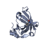
- Assembly
| Deposited unit | 
| ||||||||||
|---|---|---|---|---|---|---|---|---|---|---|---|
| 1 | 
| ||||||||||
| 2 | 
| ||||||||||
| Unit cell |
| ||||||||||
| Components on special symmetry positions |
|
-Components
-Protein / DNA chain , 2 types, 4 molecules CADB
| #1: Protein | Mass: 12385.729 Da / Num. of mol.: 2 / Mutation: Y91F Source method: isolated from a genetically manipulated source Source: (gene. exp.) Muscovy duck circovirus / Production host:  #2: DNA chain | Mass: 2993.990 Da / Num. of mol.: 2 / Source method: obtained synthetically / Source: (synth.) Muscovy duck circovirus |
|---|
-Non-polymers , 4 types, 287 molecules 


| #3: Chemical | ChemComp-GOL /  Mass: 92.094 Da / Num. of mol.: 1 / Source method: obtained synthetically / Formula: C3H8O3 Mass: 92.094 Da / Num. of mol.: 1 / Source method: obtained synthetically / Formula: C3H8O3 | ||||
|---|---|---|---|---|---|
| #4: Chemical | ChemComp-SO4 /  Mass: 96.063 Da / Num. of mol.: 4 / Source method: obtained synthetically / Formula: SO4 Mass: 96.063 Da / Num. of mol.: 4 / Source method: obtained synthetically / Formula: SO4#5: Chemical | ChemComp-MN / |  Mass: 54.938 Da / Num. of mol.: 1 / Source method: obtained synthetically / Formula: Mn Mass: 54.938 Da / Num. of mol.: 1 / Source method: obtained synthetically / Formula: Mn#6: Water | ChemComp-HOH / | Mass: 18.015 Da / Num. of mol.: 281 / Source method: isolated from a natural source / Formula: H2O |
-Details
| Has ligand of interest | N |
|---|
-Experimental details
-Experiment
| Experiment | Method: X-RAY DIFFRACTION / Number of used crystals: 1 |
|---|
- Sample preparation
| Crystal | Density Matthews: 3.44 Å3/Da / Density % sol: 64.26 % |
|---|---|
| Crystal grow | Temperature: 298 K / Method: vapor diffusion, hanging drop / pH: 4.6 Details: 0.1 M sodium acetate, pH 4.8, 2.2M ammonium sulfate, 20% glycerol added to drop |
-Data collection
| Diffraction | Mean temperature: 100 K / Serial crystal experiment: N |
|---|---|
| Diffraction source | Source: SYNCHROTRON / Site: APS / Beamline: 24-ID-C / Wavelength: 0.9791 Å |
| Detector | Type: DECTRIS PILATUS3 S 6M / Detector: PIXEL / Date: Sep 27, 2020 |
| Radiation | Protocol: SINGLE WAVELENGTH / Monochromatic (M) / Laue (L): M / Scattering type: x-ray |
| Radiation wavelength | Wavelength: 0.9791 Å / Relative weight: 1 |
| Reflection | Resolution: 1.69→54.65 Å / Num. obs: 48935 / % possible obs: 99.84 % / Redundancy: 8.7 % / Biso Wilson estimate: 28.27 Å2 / CC1/2: 1 / CC star: 1 / Rmerge(I) obs: 0.03628 / Rpim(I) all: 0.01275 / Rrim(I) all: 0.03856 / Net I/σ(I): 32.89 |
| Reflection shell | Resolution: 1.69→1.751 Å / Redundancy: 8.5 % / Rmerge(I) obs: 0.8253 / Mean I/σ(I) obs: 2.28 / Num. unique obs: 4791 / CC1/2: 0.784 / CC star: 0.938 / Rpim(I) all: 0.2922 / Rrim(I) all: 0.8776 / % possible all: 99.69 |
- Processing
| Software |
| ||||||||||||||||||||||||||||||||||||||||||||||||||||||||||||||||||||||||||||||||||||||||||||||||||||||||||||||||||||||||||||||
|---|---|---|---|---|---|---|---|---|---|---|---|---|---|---|---|---|---|---|---|---|---|---|---|---|---|---|---|---|---|---|---|---|---|---|---|---|---|---|---|---|---|---|---|---|---|---|---|---|---|---|---|---|---|---|---|---|---|---|---|---|---|---|---|---|---|---|---|---|---|---|---|---|---|---|---|---|---|---|---|---|---|---|---|---|---|---|---|---|---|---|---|---|---|---|---|---|---|---|---|---|---|---|---|---|---|---|---|---|---|---|---|---|---|---|---|---|---|---|---|---|---|---|---|---|---|---|---|
| Refinement | Method to determine structure: MOLECULAR REPLACEMENT Starting model: 6WDZ Resolution: 1.69→54.65 Å / SU ML: 0.27 / Cross valid method: THROUGHOUT / σ(F): 1.34 / Phase error: 17.5 / Stereochemistry target values: ML
| ||||||||||||||||||||||||||||||||||||||||||||||||||||||||||||||||||||||||||||||||||||||||||||||||||||||||||||||||||||||||||||||
| Solvent computation | Shrinkage radii: 0.9 Å / VDW probe radii: 1.11 Å / Solvent model: FLAT BULK SOLVENT MODEL | ||||||||||||||||||||||||||||||||||||||||||||||||||||||||||||||||||||||||||||||||||||||||||||||||||||||||||||||||||||||||||||||
| Displacement parameters | Biso max: 87.21 Å2 / Biso mean: 34.3287 Å2 / Biso min: 20.12 Å2 | ||||||||||||||||||||||||||||||||||||||||||||||||||||||||||||||||||||||||||||||||||||||||||||||||||||||||||||||||||||||||||||||
| Refinement step | Cycle: final / Resolution: 1.69→54.65 Å
| ||||||||||||||||||||||||||||||||||||||||||||||||||||||||||||||||||||||||||||||||||||||||||||||||||||||||||||||||||||||||||||||
| LS refinement shell | Refine-ID: X-RAY DIFFRACTION / Rfactor Rfree error: 0 / Total num. of bins used: 17
|
