 Movie
Movie Controller

[English] 日本語
 Yorodumi
- PDB-7kh7: Crystal structure of Staphylococcus aureus ketol-acid reductoisom... -
+ Open data

- Basic information
| Entry | Database: PDB / ID: 7kh7 | ||||||
|---|---|---|---|---|---|---|---|
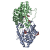
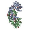
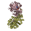
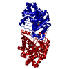
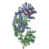
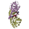
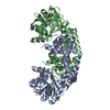
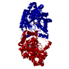
| Title | Crystal structure of Staphylococcus aureus ketol-acid reductoisomerase in complex with Mg2+, NADPH, and NSC116565 | ||||||
 Components Components | Ketol-acid reductoisomerase (NADP(+)) | ||||||
 Keywords Keywords | ISOMERASE/Inhibitor / REDUCTOISOMERASE / INHIBITOR / COMPLEX / METAL BINDING PROTEIN / ISOMERASE / ISOMERASE-Inhibitor complex | ||||||
| Function / homology |  Function and homology information Function and homology informationketol-acid reductoisomerase (NADP+) / ketol-acid reductoisomerase activity / L-valine biosynthetic process / : / isomerase activity / NADP binding / magnesium ion binding / cytosol Similarity search - Function | ||||||
| Biological species |   Staphylococcus aureus (bacteria) Staphylococcus aureus (bacteria) | ||||||
| Method |  X-RAY DIFFRACTION / SYNCHROTRON / MOLECULAR REPLACEMENT / Resolution: 2.63 Å X-RAY DIFFRACTION / SYNCHROTRON / MOLECULAR REPLACEMENT / Resolution: 2.63 Å | ||||||
 Authors Authors | Kurz, J.L. / Patel, K.P. / Guddat, L.W. | ||||||
| Funding support |  Australia, 1items Australia, 1items
| ||||||
 Citation Citation | Journal: Chemistry / Year: 2021 Title: Discovery of a Pyrimidinedione Derivative with Potent Inhibitory Activity against Mycobacterium tuberculosis Ketol-Acid Reductoisomerase. Authors: Lin, X. / Kurz, J.L. / Patel, K.M. / Wun, S.J. / Hussein, W.M. / Lonhienne, T. / West, N.P. / McGeary, R.P. / Schenk, G. / Guddat, L.W. | ||||||
| History |
|
- Structure visualization
| Structure viewer | Molecule: MolmilJmol/JSmol |
|---|
- Downloads & links
-Download
| PDBx/mmCIF format | 7kh7.cif.gz | 154.6 KB | Display | PDBx/mmCIF format |
|---|---|---|---|---|
| PDB format | pdb7kh7.ent.gz | 109.2 KB | Display | PDB format |
| PDBx/mmJSON format | 7kh7.json.gz | Tree view | PDBx/mmJSON format | |
| Others |  Other downloads Other downloads |
-Validation report
| Arichive directory | https://data.pdbj.org/pub/pdb/validation_reports/kh/7kh7ftp://data.pdbj.org/pub/pdb/validation_reports/kh/7kh7 | HTTPS FTP |
|---|
-Related structure data
| Related structure data |  7ke2C 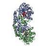 5w3kS S: Starting model for refinement C: citing same article ( |
|---|---|
| Similar structure data |


-Links
 PDBj
PDBj


- Assembly
| Deposited unit | 
| ||||||||||||
|---|---|---|---|---|---|---|---|---|---|---|---|---|---|
| 1 |
| ||||||||||||
| Unit cell |
|
-Components
-Protein , 1 types, 2 molecules AB
| #1: Protein | Mass: 37886.645 Da / Num. of mol.: 2 Source method: isolated from a genetically manipulated source Source: (gene. exp.) Staphylococcus aureus (bacteria) / Gene: ilvC, BMF23_13825, BO217_1422, ERS072840_02559 / Production host: References: UniProt: A0A145BYP4, ketol-acid reductoisomerase (NADP+) |
|---|
-Non-polymers , 5 types, 70 molecules 


| #2: Chemical | ChemComp-NAP /  Mass: 743.405 Da / Num. of mol.: 1 / Source method: obtained synthetically / Formula: C21H28N7O17P3 Mass: 743.405 Da / Num. of mol.: 1 / Source method: obtained synthetically / Formula: C21H28N7O17P3 | ||||
|---|---|---|---|---|---|
| #3: Chemical | ChemComp-NDP /  Mass: 745.421 Da / Num. of mol.: 1 / Source method: obtained synthetically / Formula: C21H30N7O17P3 Mass: 745.421 Da / Num. of mol.: 1 / Source method: obtained synthetically / Formula: C21H30N7O17P3 | ||||
| #4: Chemical | ChemComp-MG /  Mass: 24.305 Da / Num. of mol.: 5 / Source method: obtained synthetically / Formula: Mg Mass: 24.305 Da / Num. of mol.: 5 / Source method: obtained synthetically / Formula: Mg#5: Chemical |  Mass: 199.187 Da / Num. of mol.: 2 / Source method: obtained synthetically / Formula: C6H5N3O3S / Feature type: SUBJECT OF INVESTIGATION Mass: 199.187 Da / Num. of mol.: 2 / Source method: obtained synthetically / Formula: C6H5N3O3S / Feature type: SUBJECT OF INVESTIGATION#6: Water | ChemComp-HOH / | Mass: 18.015 Da / Num. of mol.: 61 / Source method: isolated from a natural source / Formula: H2O |
-Details
| Has ligand of interest | Y |
|---|
-Experimental details
-Experiment
| Experiment | Method: X-RAY DIFFRACTION / Number of used crystals: 1 |
|---|
- Sample preparation
| Crystal | Density Matthews: 2.16 Å3/Da / Density % sol: 43.04 % |
|---|---|
| Crystal grow | Temperature: 289.5 K / Method: vapor diffusion, hanging drop / pH: 8.1 / Details: 0.2 M sodium acetate pH 8.1, 32.5% PEG3350 |
-Data collection
| Diffraction | Mean temperature: 100 K / Serial crystal experiment: N |
|---|---|
| Diffraction source | Source: SYNCHROTRON / Site: Australian Synchrotron / Beamline: MX2 / Wavelength: 0.953 Å |
| Detector | Type: DECTRIS EIGER2 X 1M / Detector: PIXEL / Date: Nov 1, 2019 |
| Radiation | Monochromator: M / Protocol: SINGLE WAVELENGTH / Monochromatic (M) / Laue (L): M / Scattering type: x-ray |
| Radiation wavelength | Wavelength: 0.953 Å / Relative weight: 1 |
| Reflection | Resolution: 2.62→48.84 Å / Num. obs: 20203 / % possible obs: 99.6 % / Redundancy: 13.44 % / Biso Wilson estimate: 54.4 Å2 / Rpim(I) all: 0.05 / Net I/σ(I): 9.7 |
| Reflection shell | Resolution: 2.62→2.74 Å / Redundancy: 15.7 % / Rmerge(I) obs: 0.51 / Mean I/σ(I) obs: 1.4 / Num. unique obs: 2352 / Rpim(I) all: 0.511 / % possible all: 96.8 |
- Processing
| Software |
| |||||||||||||||||||||||||||||||||||||||||||||||||||||||||||||||||||||||||||||||||||||||||||||||||||||||||
|---|---|---|---|---|---|---|---|---|---|---|---|---|---|---|---|---|---|---|---|---|---|---|---|---|---|---|---|---|---|---|---|---|---|---|---|---|---|---|---|---|---|---|---|---|---|---|---|---|---|---|---|---|---|---|---|---|---|---|---|---|---|---|---|---|---|---|---|---|---|---|---|---|---|---|---|---|---|---|---|---|---|---|---|---|---|---|---|---|---|---|---|---|---|---|---|---|---|---|---|---|---|---|---|---|---|---|
| Refinement | Method to determine structure: MOLECULAR REPLACEMENT Starting model: 5w3k Resolution: 2.63→48.84 Å / SU ML: 0.3715 / Cross valid method: FREE R-VALUE / σ(F): 1.35 / Phase error: 35.5494 Stereochemistry target values: GeoStd + Monomer Library + CDL v1.2
| |||||||||||||||||||||||||||||||||||||||||||||||||||||||||||||||||||||||||||||||||||||||||||||||||||||||||
| Solvent computation | Shrinkage radii: 0.9 Å / VDW probe radii: 1.11 Å / Solvent model: FLAT BULK SOLVENT MODEL | |||||||||||||||||||||||||||||||||||||||||||||||||||||||||||||||||||||||||||||||||||||||||||||||||||||||||
| Displacement parameters | Biso mean: 63.1 Å2 | |||||||||||||||||||||||||||||||||||||||||||||||||||||||||||||||||||||||||||||||||||||||||||||||||||||||||
| Refinement step | Cycle: LAST / Resolution: 2.63→48.84 Å
| |||||||||||||||||||||||||||||||||||||||||||||||||||||||||||||||||||||||||||||||||||||||||||||||||||||||||
| Refine LS restraints |
| |||||||||||||||||||||||||||||||||||||||||||||||||||||||||||||||||||||||||||||||||||||||||||||||||||||||||
| LS refinement shell |
|
