 Movie
Movie Controller


+ Open data

- Basic information
| Entry | Database: PDB / ID: 7kgz | ||||||
|---|---|---|---|---|---|---|---|
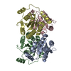
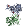
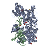
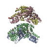
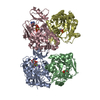
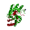
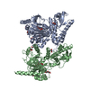
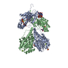
| Title | FMN-binding beta-glucuronidase from Roseburia hominis | ||||||
 Components Components | Beta-glucuronidase | ||||||
 Keywords Keywords | HYDROLASE / Beta-glucuronidase / gut microbial enzyme / FMN | ||||||
| Function / homology | FLAVIN MONONUCLEOTIDE Function and homology information Function and homology information | ||||||
| Biological species |  Roseburia hominis (bacteria) Roseburia hominis (bacteria) | ||||||
| Method |  X-RAY DIFFRACTION / SYNCHROTRON / MOLECULAR REPLACEMENT / Resolution: 2.4 Å X-RAY DIFFRACTION / SYNCHROTRON / MOLECULAR REPLACEMENT / Resolution: 2.4 Å | ||||||
 Authors Authors | Walker, M.E. / Redinbo, M.R. | ||||||
| Funding support |  United States, 1items United States, 1items
| ||||||
 Citation Citation | Journal: Nat Commun / Year: 2022 Title: Microbial enzymes induce colitis by reactivating triclosan in the mouse gastrointestinal tract. Authors: Zhang, J. / Walker, M.E. / Sanidad, K.Z. / Zhang, H. / Liang, Y. / Zhao, E. / Chacon-Vargas, K. / Yeliseyev, V. / Parsonnet, J. / Haggerty, T.D. / Wang, G. / Simpson, J.B. / Jariwala, P.B. / ...Authors: Zhang, J. / Walker, M.E. / Sanidad, K.Z. / Zhang, H. / Liang, Y. / Zhao, E. / Chacon-Vargas, K. / Yeliseyev, V. / Parsonnet, J. / Haggerty, T.D. / Wang, G. / Simpson, J.B. / Jariwala, P.B. / Beaty, V.V. / Yang, J. / Yang, H. / Panigrahy, A. / Minter, L.M. / Kim, D. / Gibbons, J.G. / Liu, L. / Li, Z. / Xiao, H. / Borlandelli, V. / Overkleeft, H.S. / Cloer, E.W. / Major, M.B. / Goldfarb, D. / Cai, Z. / Redinbo, M.R. / Zhang, G. | ||||||
| History |
|
- Structure visualization
| Structure viewer | Molecule: MolmilJmol/JSmol |
|---|
- Downloads & links
-Download
| PDBx/mmCIF format | 7kgz.cif.gz | 351.9 KB | Display | PDBx/mmCIF format |
|---|---|---|---|---|
| PDB format | pdb7kgz.ent.gz | 224.9 KB | Display | PDB format |
| PDBx/mmJSON format | 7kgz.json.gz | Tree view | PDBx/mmJSON format | |
| Others |  Other downloads Other downloads |
-Validation report
| Arichive directory | https://data.pdbj.org/pub/pdb/validation_reports/kg/7kgzftp://data.pdbj.org/pub/pdb/validation_reports/kg/7kgz | HTTPS FTP |
|---|
-Related structure data
| Related structure data |  7kgyC  6mvhS S: Starting model for refinement C: citing same article ( |
|---|---|
| Similar structure data |
-Links
 PDBj
PDBj- Assembly
| Deposited unit | 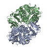
| ||||||||||||
|---|---|---|---|---|---|---|---|---|---|---|---|---|---|
| 1 |
| ||||||||||||
| Unit cell |
|
-Components
| #1: Protein | Mass: 83204.758 Da / Num. of mol.: 2 Source method: isolated from a genetically manipulated source Source: (gene. exp.) Roseburia hominis (bacteria) / Production host: #2: Chemical |   Mass: 40.078 Da / Num. of mol.: 2 / Source method: obtained synthetically / Formula: Ca Mass: 40.078 Da / Num. of mol.: 2 / Source method: obtained synthetically / Formula: Ca#3: Chemical |   Mass: 456.344 Da / Num. of mol.: 2 / Source method: obtained synthetically / Formula: C17H21N4O9P Mass: 456.344 Da / Num. of mol.: 2 / Source method: obtained synthetically / Formula: C17H21N4O9P#4: Chemical | ChemComp-GOL /   Mass: 92.094 Da / Num. of mol.: 4 / Source method: obtained synthetically / Formula: C3H8O3 Mass: 92.094 Da / Num. of mol.: 4 / Source method: obtained synthetically / Formula: C3H8O3#5: Water | ChemComp-HOH / |  Mass: 18.015 Da / Num. of mol.: 835 / Source method: isolated from a natural source / Formula: H2O Mass: 18.015 Da / Num. of mol.: 835 / Source method: isolated from a natural source / Formula: H2OHas ligand of interest | N | |
|---|
-Experimental details
-Experiment
| Experiment | Method: X-RAY DIFFRACTION / Number of used crystals: 1 |
|---|
- Sample preparation
| Crystal | Density Matthews: 2.19 Å3/Da / Density % sol: 43.72 % |
|---|---|
| Crystal grow | Temperature: 293 K / Method: vapor diffusion, sitting drop Details: 12.5 mg/mL protein, 0.2 M lithium chloride, 20% (w/v) PEG 3350 |
-Data collection
| Diffraction | Mean temperature: 100 K / Serial crystal experiment: N |
|---|---|
| Diffraction source | Source: SYNCHROTRON / Site: APS / Beamline: 23-ID-D / Wavelength: 1.033 Å |
| Detector | Type: DECTRIS PILATUS3 6M / Detector: PIXEL / Date: Jul 26, 2019 |
| Radiation | Monochromator: Double crystal cryo-cooled Si(111) / Protocol: SINGLE WAVELENGTH / Monochromatic (M) / Laue (L): M / Scattering type: x-ray |
| Radiation wavelength | Wavelength: 1.033 Å / Relative weight: 1 |
| Reflection | Resolution: 2.4→39.07 Å / Num. obs: 55112 / % possible obs: 98.9 % / Redundancy: 3.5 % / Biso Wilson estimate: 23 Å2 / CC1/2: 0.997 / CC star: 0.999 / Rmerge(I) obs: 0.0504 / Rpim(I) all: 0.03193 / Rrim(I) all: 0.0598 / Net I/σ(I): 18.68 |
| Reflection shell | Resolution: 2.4→2.486 Å / Redundancy: 3.5 % / Rmerge(I) obs: 0.1243 / Mean I/σ(I) obs: 8.37 / Num. unique obs: 5470 / CC1/2: 0.981 / CC star: 0.995 / Rpim(I) all: 0.0781 / Rrim(I) all: 0.147 / % possible all: 98.38 |
- Processing
| Software |
| |||||||||||||||||||||||||||||||||||||||||||||||||||||||||||||||||||||||||||||||||||||||||||||||||||||||||
|---|---|---|---|---|---|---|---|---|---|---|---|---|---|---|---|---|---|---|---|---|---|---|---|---|---|---|---|---|---|---|---|---|---|---|---|---|---|---|---|---|---|---|---|---|---|---|---|---|---|---|---|---|---|---|---|---|---|---|---|---|---|---|---|---|---|---|---|---|---|---|---|---|---|---|---|---|---|---|---|---|---|---|---|---|---|---|---|---|---|---|---|---|---|---|---|---|---|---|---|---|---|---|---|---|---|---|
| Refinement | Method to determine structure: MOLECULAR REPLACEMENT Starting model: 6MVH Resolution: 2.4→39.07 Å / SU ML: 0.2251 / Cross valid method: FREE R-VALUE / σ(F): 1.34 / Phase error: 19.9284 Stereochemistry target values: GeoStd + Monomer Library + CDL v1.2
| |||||||||||||||||||||||||||||||||||||||||||||||||||||||||||||||||||||||||||||||||||||||||||||||||||||||||
| Solvent computation | Shrinkage radii: 0.9 Å / VDW probe radii: 1.11 Å / Solvent model: FLAT BULK SOLVENT MODEL | |||||||||||||||||||||||||||||||||||||||||||||||||||||||||||||||||||||||||||||||||||||||||||||||||||||||||
| Displacement parameters | Biso mean: 24.73 Å2 | |||||||||||||||||||||||||||||||||||||||||||||||||||||||||||||||||||||||||||||||||||||||||||||||||||||||||
| Refinement step | Cycle: LAST / Resolution: 2.4→39.07 Å
| |||||||||||||||||||||||||||||||||||||||||||||||||||||||||||||||||||||||||||||||||||||||||||||||||||||||||
| Refine LS restraints |
| |||||||||||||||||||||||||||||||||||||||||||||||||||||||||||||||||||||||||||||||||||||||||||||||||||||||||
| LS refinement shell |
|
