 Movie
Movie Controller

[English] 日本語
 Yorodumi
- PDB-7kgq: Crystal Structure of HLA-A*0201in complex with SARS-CoV-2 N222-230 -
+ Open data

- Basic information
| Entry | Database: PDB / ID: 7kgq | |||||||||
|---|---|---|---|---|---|---|---|---|---|---|
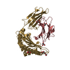
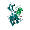
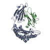
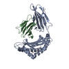
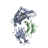
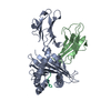
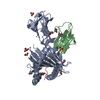
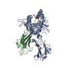
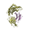
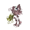
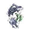
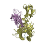
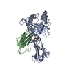
| Title | Crystal Structure of HLA-A*0201in complex with SARS-CoV-2 N222-230 | |||||||||
 Components Components |
| |||||||||
 Keywords Keywords | IMMUNE SYSTEM / HLA-A*0201 / T cell / SARS-CoV-2 / COVID-19 / viral peptide / TCR | |||||||||
| Function / homology |  Function and homology information Function and homology informationresponse to host immune response / viral RNA genome packaging / negative regulation of interferon-beta production / Maturation of nucleoprotein / poly(U) RNA binding / positive regulation of NLRP3 inflammasome complex assembly / MHC class I protein binding / CD28 dependent PI3K/Akt signaling / antigen processing and presentation of peptide antigen via MHC class I / SARS-CoV-2 targets host intracellular signalling and regulatory pathways ...response to host immune response / viral RNA genome packaging / negative regulation of interferon-beta production / Maturation of nucleoprotein / poly(U) RNA binding / positive regulation of NLRP3 inflammasome complex assembly / MHC class I protein binding / CD28 dependent PI3K/Akt signaling / antigen processing and presentation of peptide antigen via MHC class I / SARS-CoV-2 targets host intracellular signalling and regulatory pathways / early endosome lumen / Nef mediated downregulation of MHC class I complex cell surface expression / DAP12 interactions / T cell mediated cytotoxicity / Endosomal/Vacuolar pathway / VEGFR2 mediated vascular permeability / Antigen Presentation: Folding, assembly and peptide loading of class I MHC / lumenal side of endoplasmic reticulum membrane / regulation of iron ion transport / cellular response to iron(III) ion / antigen processing and presentation of exogenous protein antigen via MHC class Ib, TAP-dependent / negative regulation of iron ion transport / negative regulation of forebrain neuron differentiation / regulation of erythrocyte differentiation / peptide antigen assembly with MHC class I protein complex / protein sequestering activity / ER to Golgi transport vesicle membrane / response to molecule of bacterial origin / molecular condensate scaffold activity / HFE-transferrin receptor complex / MHC class I peptide loading complex / transferrin transport / negative regulation of receptor-mediated endocytosis / cellular response to iron ion / positive regulation of T cell cytokine production / antigen processing and presentation of endogenous peptide antigen via MHC class I / MHC class I protein complex / peptide antigen assembly with MHC class II protein complex / NOD1/2 Signaling Pathway / TAK1-dependent IKK and NF-kappa-B activation / negative regulation of neurogenesis / multicellular organismal-level iron ion homeostasis / cellular response to nicotine / MHC class II protein complex / positive regulation of receptor-mediated endocytosis / positive regulation of T cell mediated cytotoxicity / DDX58/IFIH1-mediated induction of interferon-alpha/beta / RNA stem-loop binding / negative regulation of epithelial cell proliferation / specific granule lumen / antigen processing and presentation of exogenous peptide antigen via MHC class II / positive regulation of immune response / peptide antigen binding / phagocytic vesicle membrane / recycling endosome membrane / positive regulation of T cell activation / Interferon gamma signaling / Immunoregulatory interactions between a Lymphoid and a non-Lymphoid cell / Interleukin-1 signaling / viral capsid / Interferon alpha/beta signaling / Modulation by Mtb of host immune system / sensory perception of smell / positive regulation of cellular senescence / tertiary granule lumen / MHC class II protein complex binding / T cell differentiation in thymus / DAP12 signaling / PIP3 activates AKT signaling / late endosome membrane / negative regulation of neuron projection development / protein refolding / viral nucleocapsid / ER-Phagosome pathway / Transcription of SARS-CoV-2 sgRNAs / early endosome membrane / Translation of Structural Proteins / Virion Assembly and Release / host cell endoplasmic reticulum-Golgi intermediate compartment / host extracellular region / Induction of Cell-Cell Fusion / amyloid fibril formation / protein homotetramerization / intracellular iron ion homeostasis / host cell Golgi apparatus / Attachment and Entry / learning or memory / immune response / host cell perinuclear region of cytoplasm / endoplasmic reticulum lumen / ribonucleoprotein complex / Amyloid fiber formation / Golgi membrane / external side of plasma membrane / lysosomal membrane / focal adhesion / Neutrophil degranulation / SARS-CoV-2 activates/modulates innate and adaptive immune responses / structural molecule activity / Golgi apparatus Similarity search - Function | |||||||||
| Biological species |  Homo sapiens (human) Homo sapiens (human)  Severe acute respiratory syndrome coronavirus 2 Severe acute respiratory syndrome coronavirus 2 | |||||||||
| Method |  X-RAY DIFFRACTION / SYNCHROTRON / MOLECULAR REPLACEMENT / molecular replacement / Resolution: 1.34 Å X-RAY DIFFRACTION / SYNCHROTRON / MOLECULAR REPLACEMENT / molecular replacement / Resolution: 1.34 Å | |||||||||
 Authors Authors | Szeto, C. / Chatzileontiadou, D.S.M. / Riboldi-Tunnicliffe, A. / Gras, S. | |||||||||
| Funding support |  Australia, 2items Australia, 2items
| |||||||||
 Citation Citation | Journal: Iscience / Year: 2021 Title: The presentation of SARS-CoV-2 peptides by the common HLA-A * 02:01 molecule. Authors: Szeto, C. / Chatzileontiadou, D.S.M. / Nguyen, A.T. / Sloane, H. / Lobos, C.A. / Jayasinghe, D. / Halim, H. / Smith, C. / Riboldi-Tunnicliffe, A. / Grant, E.J. / Gras, S. | |||||||||
| History |
|
- Structure visualization
| Structure viewer | Molecule: MolmilJmol/JSmol |
|---|
- Downloads & links
-Download
| PDBx/mmCIF format | 7kgq.cif.gz | 112.8 KB | Display | PDBx/mmCIF format |
|---|---|---|---|---|
| PDB format | pdb7kgq.ent.gz | 83.2 KB | Display | PDB format |
| PDBx/mmJSON format | 7kgq.json.gz | Tree view | PDBx/mmJSON format | |
| Others |  Other downloads Other downloads |
-Validation report
| Arichive directory | https://data.pdbj.org/pub/pdb/validation_reports/kg/7kgqftp://data.pdbj.org/pub/pdb/validation_reports/kg/7kgq | HTTPS FTP |
|---|
-Related structure data
| Related structure data |  7kgoC  7kgpC  7kgrC  7kgsC  7kgtC  3gsoS S: Starting model for refinement C: citing same article ( |
|---|---|
| Similar structure data |


-Links
 PDBj
PDBj


- Assembly
| Deposited unit | 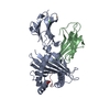
| ||||||||
|---|---|---|---|---|---|---|---|---|---|
| 1 |
| ||||||||
| Unit cell |
|
-Components
-Protein , 2 types, 2 molecules AB
| #1: Protein | Mass: 32153.523 Da / Num. of mol.: 1 Source method: isolated from a genetically manipulated source Source: (gene. exp.) Homo sapiens (human) / Gene: HLA-A / Plasmid: pET / Production host:  |
|---|---|
| #2: Protein | Mass: 11748.160 Da / Num. of mol.: 1 Source method: isolated from a genetically manipulated source Source: (gene. exp.) Homo sapiens (human) / Gene: B2M, CDABP0092, HDCMA22P / Plasmid: pET / Production host: |
-Protein/peptide , 1 types, 1 molecules C
| #3: Protein/peptide | Mass: 1098.317 Da / Num. of mol.: 1 / Fragment: residues 222-230 / Source method: obtained synthetically / Details: synthesized Source: (synth.) Severe acute respiratory syndrome coronavirus 2References: UniProt: P0DTC9 |
|---|
-Non-polymers , 4 types, 599 molecules 


| #4: Chemical |  Mass: 112.411 Da / Num. of mol.: 2 / Source method: obtained synthetically / Formula: Cd Mass: 112.411 Da / Num. of mol.: 2 / Source method: obtained synthetically / Formula: Cd#5: Chemical |  Mass: 40.078 Da / Num. of mol.: 2 / Source method: obtained synthetically / Formula: Ca Mass: 40.078 Da / Num. of mol.: 2 / Source method: obtained synthetically / Formula: Ca#6: Chemical | ChemComp-EDO / |  Mass: 62.068 Da / Num. of mol.: 1 / Source method: obtained synthetically / Formula: C2H6O2 Mass: 62.068 Da / Num. of mol.: 1 / Source method: obtained synthetically / Formula: C2H6O2#7: Water | ChemComp-HOH / | Mass: 18.015 Da / Num. of mol.: 594 / Source method: isolated from a natural source / Formula: H2O |
|---|
-Details
| Has ligand of interest | N |
|---|---|
| Has protein modification | Y |
-Experimental details
-Experiment
| Experiment | Method: X-RAY DIFFRACTION / Number of used crystals: 1 |
|---|
- Sample preparation
| Crystal | Density Matthews: 2.97 Å3/Da / Density % sol: 58.58 % / Mosaicity: 0.05 ° |
|---|---|
| Crystal grow | Temperature: 277 K / Method: vapor diffusion, hanging drop / pH: 8 / Details: 20% PEG3350, 0.2M KFormate, 1mM CaCl2 |
-Data collection
| Diffraction | Mean temperature: 100 K / Serial crystal experiment: N | ||||||||||||||||||||||||||||||
|---|---|---|---|---|---|---|---|---|---|---|---|---|---|---|---|---|---|---|---|---|---|---|---|---|---|---|---|---|---|---|---|
| Diffraction source | Source: SYNCHROTRON / Site: Australian Synchrotron / Beamline: MX2 / Wavelength: 0.95369 Å | ||||||||||||||||||||||||||||||
| Detector | Type: DECTRIS EIGER X 16M / Detector: PIXEL / Date: Jun 6, 2020 | ||||||||||||||||||||||||||||||
| Radiation | Protocol: SINGLE WAVELENGTH / Monochromatic (M) / Laue (L): M / Scattering type: x-ray | ||||||||||||||||||||||||||||||
| Radiation wavelength | Wavelength: 0.95369 Å / Relative weight: 1 | ||||||||||||||||||||||||||||||
| Reflection | Resolution: 1.34→48.02 Å / Num. obs: 120760 / % possible obs: 99.9 % / Redundancy: 13.4 % / Biso Wilson estimate: 17.37 Å2 / CC1/2: 0.999 / Rmerge(I) obs: 0.08 / Rpim(I) all: 0.023 / Rrim(I) all: 0.083 / Net I/σ(I): 17.1 / Num. measured all: 1621917 / Scaling rejects: 6 | ||||||||||||||||||||||||||||||
| Reflection shell | Diffraction-ID: 1
|
-Phasing
| Phasing | Method: molecular replacement |
|---|
- Processing
| Software |
| ||||||||||||||||||||||||||||||||||||||||||||||||||||||||||||||||||||||||||||||||||||||||||||||||||||||||||||
|---|---|---|---|---|---|---|---|---|---|---|---|---|---|---|---|---|---|---|---|---|---|---|---|---|---|---|---|---|---|---|---|---|---|---|---|---|---|---|---|---|---|---|---|---|---|---|---|---|---|---|---|---|---|---|---|---|---|---|---|---|---|---|---|---|---|---|---|---|---|---|---|---|---|---|---|---|---|---|---|---|---|---|---|---|---|---|---|---|---|---|---|---|---|---|---|---|---|---|---|---|---|---|---|---|---|---|---|---|---|
| Refinement | Method to determine structure: MOLECULAR REPLACEMENT Starting model: 3GSO Resolution: 1.34→19.6 Å / Cor.coef. Fo:Fc: 0.965 / Cor.coef. Fo:Fc free: 0.96 / SU R Cruickshank DPI: 0.046 / Cross valid method: THROUGHOUT / σ(F): 0 / SU R Blow DPI: 0.05 / SU Rfree Blow DPI: 0.051 / SU Rfree Cruickshank DPI: 0.048
| ||||||||||||||||||||||||||||||||||||||||||||||||||||||||||||||||||||||||||||||||||||||||||||||||||||||||||||
| Displacement parameters | Biso max: 101 Å2 / Biso mean: 22.34 Å2 / Biso min: 7.74 Å2
| ||||||||||||||||||||||||||||||||||||||||||||||||||||||||||||||||||||||||||||||||||||||||||||||||||||||||||||
| Refine analyze | Luzzati coordinate error obs: 0.16 Å | ||||||||||||||||||||||||||||||||||||||||||||||||||||||||||||||||||||||||||||||||||||||||||||||||||||||||||||
| Refinement step | Cycle: final / Resolution: 1.34→19.6 Å
| ||||||||||||||||||||||||||||||||||||||||||||||||||||||||||||||||||||||||||||||||||||||||||||||||||||||||||||
| Refine LS restraints |
| ||||||||||||||||||||||||||||||||||||||||||||||||||||||||||||||||||||||||||||||||||||||||||||||||||||||||||||
| LS refinement shell | Resolution: 1.34→1.35 Å / Rfactor Rfree error: 0 / Total num. of bins used: 50
|
