 Movie
Movie Controller

[English] 日本語
 Yorodumi
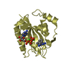
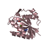
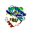
- PDB-7kdo: Crystal structure of Escherichia coli HPPK in complex with bisubs... -
+ Open data

- Basic information
| Entry | Database: PDB / ID: 7kdo | ||||||
|---|---|---|---|---|---|---|---|
| Title | Crystal structure of Escherichia coli HPPK in complex with bisubstrate inhibitor HP-73 | ||||||
 Components Components | 2-amino-4-hydroxy-6-hydroxymethyldihydropteridine pyrophosphokinase | ||||||
 Keywords Keywords | TRANSFERASE/INHIBITOR / alpha beta / TRANSFERASE / TRANSFERASE-INHIBITOR complex | ||||||
| Function / homology |  Function and homology information Function and homology information2-amino-4-hydroxy-6-hydroxymethyldihydropteridine diphosphokinase / 2-amino-4-hydroxy-6-hydroxymethyldihydropteridine diphosphokinase activity / folic acid biosynthetic process / tetrahydrofolate biosynthetic process / kinase activity / magnesium ion binding / ATP binding Similarity search - Function | ||||||
| Biological species |  | ||||||
| Method |  X-RAY DIFFRACTION / SYNCHROTRON / FOURIER SYNTHESIS / Resolution: 1.6 Å X-RAY DIFFRACTION / SYNCHROTRON / FOURIER SYNTHESIS / Resolution: 1.6 Å | ||||||
 Authors Authors | Shaw, G.X. / Shi, G. / Ji, X. | ||||||
 Citation Citation | Journal: Bioorg.Med.Chem. / Year: 2021 Title: Bisubstrate inhibitors of 6-hydroxymethyl-7,8-dihydropterin pyrophosphokinase: Transition state analogs for high affinity binding. Authors: Shi, G. / Shaw, G.X. / Zhu, F. / Tarasov, S.G. / Ji, X. #1: Journal: J. Med. Chem. / Year: 2001Title: Bisubstrate analogue inhibitors of 6-hydroxymethyl-7,8-dihydropterin pyrophosphokinase: Synthesis and biochemical and crystallographic studies Authors: Shi, G. / Blaszczyk, J. / Ji, X. / Yan, H. #2: Journal: Bioorg. Med. Chem. / Year: 2012Title: Bisubstrate analogue inhibitors of 6-hydroxymethyl-7,8-dihydropterin pyrophosphokinase: New design with improved properties Authors: Shi, G. / Shaw, G. / Liang, Y.-H. / Subburamann, P. / Li, Y. / Wu, Y. / Yan, H. / Ji, X. #3: Journal: Bioorg. Med. Chem. / Year: 2012Title: Bisubstrate analog inhibitors of 6-hydroxymethyl-7,8-dihydropterin pyrophosphokinase: New lead exhibits a distinct binding mode Authors: Shi, G. / Shaw, G. / Li, Y. / Wu, Y. / Yan, H. / Ji, X. #4: Journal: FEBS J. / Year: 2014Title: Structural enzymology and inhibition of the bi-functional folate pathway enzyme HPPK-DHPS from the biowarfare agent Francisella tularensis Authors: Shaw, G.X. / Li, Y. / Shi, G. / Wu, Y. / Cherry, S. / Needle, D. / Zhang, D. / Tropea, J.E. / Waugh, D.S. / Yan, H. / Ji, X. | ||||||
| History |
|
- Structure visualization
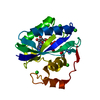
| Structure viewer | Molecule: MolmilJmol/JSmol |
|---|
- Downloads & links
-Download
| PDBx/mmCIF format | 7kdo.cif.gz | 86.3 KB | Display | PDBx/mmCIF format |
|---|---|---|---|---|
| PDB format | pdb7kdo.ent.gz | 59.7 KB | Display | PDB format |
| PDBx/mmJSON format | 7kdo.json.gz | Tree view | PDBx/mmJSON format | |
| Others |  Other downloads Other downloads |
-Validation report
| Arichive directory | https://data.pdbj.org/pub/pdb/validation_reports/kd/7kdoftp://data.pdbj.org/pub/pdb/validation_reports/kd/7kdo | HTTPS FTP |
|---|
-Related structure data
| Related structure data | 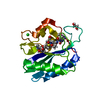 7kdrC 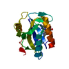 3udvS S: Starting model for refinement C: citing same article ( |
|---|---|
| Similar structure data |


-Links
 PDBj
PDBj- Assembly
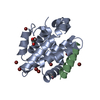
| Deposited unit | 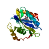
| ||||||||||||
|---|---|---|---|---|---|---|---|---|---|---|---|---|---|
| 1 |
| ||||||||||||
| Unit cell |
| ||||||||||||
| Components on special symmetry positions |
|
-Components
| #1: Protein | Mass: 17966.535 Da / Num. of mol.: 1 Source method: isolated from a genetically manipulated source Source: (gene. exp.) Strain: K12 / Gene: folK, b0142, JW0138 / Plasmid: pET17b / Production host: References: UniProt: P26281, 2-amino-4-hydroxy-6-hydroxymethyldihydropteridine diphosphokinase |
|---|---|
| #2: Chemical | ChemComp-H73 / 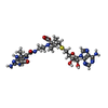  Mass: 672.716 Da / Num. of mol.: 1 / Source method: obtained synthetically / Formula: C27H36N12O7S / Feature type: SUBJECT OF INVESTIGATION Mass: 672.716 Da / Num. of mol.: 1 / Source method: obtained synthetically / Formula: C27H36N12O7S / Feature type: SUBJECT OF INVESTIGATION |
| #3: Water | ChemComp-HOH /  Mass: 18.015 Da / Num. of mol.: 170 / Source method: isolated from a natural source / Formula: H2O Mass: 18.015 Da / Num. of mol.: 170 / Source method: isolated from a natural source / Formula: H2O |
| Has ligand of interest | Y |
-Experimental details
-Experiment
| Experiment | Method: X-RAY DIFFRACTION / Number of used crystals: 1 |
|---|
- Sample preparation
| Crystal | Density Matthews: 1.88 Å3/Da / Density % sol: 34.41 % / Mosaicity: 1.526 ° / Mosaicity esd: 0.014 ° |
|---|---|
| Crystal grow | Temperature: 292 K / Method: vapor diffusion, sitting drop / pH: 7 / Details: PEG 2000 |
-Data collection
| Diffraction | Mean temperature: 100 K / Serial crystal experiment: N | |||||||||||||||||||||||||||||||||||||||||||||||||||||||||||||||||||||||||||||||||||||||||||||||||||
|---|---|---|---|---|---|---|---|---|---|---|---|---|---|---|---|---|---|---|---|---|---|---|---|---|---|---|---|---|---|---|---|---|---|---|---|---|---|---|---|---|---|---|---|---|---|---|---|---|---|---|---|---|---|---|---|---|---|---|---|---|---|---|---|---|---|---|---|---|---|---|---|---|---|---|---|---|---|---|---|---|---|---|---|---|---|---|---|---|---|---|---|---|---|---|---|---|---|---|---|---|
| Diffraction source | Source: SYNCHROTRON / Site: APS  / Beamline: 22-BM / Wavelength: 1 Å / Beamline: 22-BM / Wavelength: 1 Å | |||||||||||||||||||||||||||||||||||||||||||||||||||||||||||||||||||||||||||||||||||||||||||||||||||
| Detector | Type: RAYONIX MX300-HS / Detector: CCD / Date: Mar 27, 2015 / Details: mirrors | |||||||||||||||||||||||||||||||||||||||||||||||||||||||||||||||||||||||||||||||||||||||||||||||||||
| Radiation | Monochromator: Double crystal / Protocol: SINGLE WAVELENGTH / Monochromatic (M) / Laue (L): M / Scattering type: x-ray | |||||||||||||||||||||||||||||||||||||||||||||||||||||||||||||||||||||||||||||||||||||||||||||||||||
| Radiation wavelength | Wavelength: 1 Å / Relative weight: 1 | |||||||||||||||||||||||||||||||||||||||||||||||||||||||||||||||||||||||||||||||||||||||||||||||||||
| Reflection | Resolution: 1.6→30 Å / Num. obs: 17716 / % possible obs: 95.7 % / Redundancy: 5 % / Rmerge(I) obs: 0.067 / Rpim(I) all: 0.03 / Rrim(I) all: 0.074 / Χ2: 0.982 / Net I/σ(I): 9.6 / Num. measured all: 88152 | |||||||||||||||||||||||||||||||||||||||||||||||||||||||||||||||||||||||||||||||||||||||||||||||||||
| Reflection shell | Diffraction-ID: 1
|
- Processing
| Software |
| ||||||||||||||||||||||||||||||||||||||||||||||||||||||||
|---|---|---|---|---|---|---|---|---|---|---|---|---|---|---|---|---|---|---|---|---|---|---|---|---|---|---|---|---|---|---|---|---|---|---|---|---|---|---|---|---|---|---|---|---|---|---|---|---|---|---|---|---|---|---|---|---|---|
| Refinement | Method to determine structure: FOURIER SYNTHESIS Starting model: 3UDV Resolution: 1.6→29.93 Å / SU ML: 0.1917 / Cross valid method: FREE R-VALUE / σ(F): 1.38 / Phase error: 24.819 Stereochemistry target values: GeoStd + Monomer Library + CDL v1.2
| ||||||||||||||||||||||||||||||||||||||||||||||||||||||||
| Solvent computation | Shrinkage radii: 0.9 Å / VDW probe radii: 1.11 Å / Solvent model: FLAT BULK SOLVENT MODEL | ||||||||||||||||||||||||||||||||||||||||||||||||||||||||
| Displacement parameters | Biso mean: 25.78 Å2 | ||||||||||||||||||||||||||||||||||||||||||||||||||||||||
| Refinement step | Cycle: LAST / Resolution: 1.6→29.93 Å
| ||||||||||||||||||||||||||||||||||||||||||||||||||||||||
| Refine LS restraints |
| ||||||||||||||||||||||||||||||||||||||||||||||||||||||||
| LS refinement shell |
|
