 Movie
Movie Controller

[English] 日本語
 Yorodumi
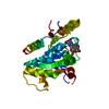
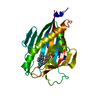
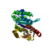
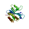
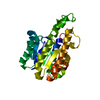
- PDB-7kcj: The crystal structure of the translation initiation factor EIF4E5... -
+ Open data

- Basic information
| Entry | Database: PDB / ID: 7kcj | ||||||
|---|---|---|---|---|---|---|---|
| Title | The crystal structure of the translation initiation factor EIF4E5 from Leishmania major | ||||||
 Components Components | Eukaryotic translation initiation factor 4E type 5 | ||||||
 Keywords Keywords | TRANSLATION | ||||||
| Function / homology |  Function and homology information Function and homology informationeukaryotic translation initiation factor 4F complex / nuclear lumen / RNA 7-methylguanosine cap binding / ciliary plasm / translation initiation factor activity / translational initiation Similarity search - Function | ||||||
| Biological species |  Leishmania major (eukaryote) Leishmania major (eukaryote) | ||||||
| Method |  X-RAY DIFFRACTION / SYNCHROTRON / MOLECULAR REPLACEMENT / Resolution: 1.997 Å X-RAY DIFFRACTION / SYNCHROTRON / MOLECULAR REPLACEMENT / Resolution: 1.997 Å | ||||||
 Authors Authors | Lima, G.B. / Guimaraes, B.G. | ||||||
| Funding support |  Brazil, 1items Brazil, 1items
| ||||||
 Citation Citation | Journal: Rna Biol. / Year: 2021 Title: The translation initiation factor EIF4E5 from Leishmania: crystal structure and interacting partners. Authors: de Lima, G.B. / de Lima Cavalcanti, T.Y.V. / de Brito, A.N.A.L.M. / de Assis, L.A. / Andrade-Vieira, R.P. / Freire, E.R. / da Silva Assuncao, T.R. / de Souza Reis, C.R. / Zanchin, N.I.T. / ...Authors: de Lima, G.B. / de Lima Cavalcanti, T.Y.V. / de Brito, A.N.A.L.M. / de Assis, L.A. / Andrade-Vieira, R.P. / Freire, E.R. / da Silva Assuncao, T.R. / de Souza Reis, C.R. / Zanchin, N.I.T. / Guimaraes, B.G. / de-Melo-Neto, O.P. | ||||||
| History |
|
- Structure visualization
| Structure viewer | Molecule: MolmilJmol/JSmol |
|---|
- Downloads & links
-Download
| PDBx/mmCIF format | 7kcj.cif.gz | 154.8 KB | Display | PDBx/mmCIF format |
|---|---|---|---|---|
| PDB format | pdb7kcj.ent.gz | 121.5 KB | Display | PDB format |
| PDBx/mmJSON format | 7kcj.json.gz | Tree view | PDBx/mmJSON format | |
| Others |  Other downloads Other downloads |
-Validation report
| Arichive directory | https://data.pdbj.org/pub/pdb/validation_reports/kc/7kcjftp://data.pdbj.org/pub/pdb/validation_reports/kc/7kcj | HTTPS FTP |
|---|
-Related structure data
| Similar structure data |
|---|


-Links
 PDBj
PDBj
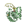
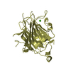
- Assembly
| Deposited unit | 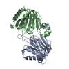
| ||||||||
|---|---|---|---|---|---|---|---|---|---|
| 1 | 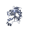
| ||||||||
| 2 | 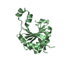
| ||||||||
| 3 |
| ||||||||
| Unit cell |
|
-Components
| #1: Protein | Mass: 25419.982 Da / Num. of mol.: 2 Source method: isolated from a genetically manipulated source Source: (gene. exp.) Leishmania major (eukaryote) / Gene: LMJF_36_0590 / Production host:  #2: Water | ChemComp-HOH / |  Mass: 18.015 Da / Num. of mol.: 204 / Source method: isolated from a natural source / Formula: H2O Mass: 18.015 Da / Num. of mol.: 204 / Source method: isolated from a natural source / Formula: H2O |
|---|
-Experimental details
-Experiment
| Experiment | Method: X-RAY DIFFRACTION / Number of used crystals: 1 |
|---|
- Sample preparation
| Crystal | Density Matthews: 1.88 Å3/Da / Density % sol: 34.63 % |
|---|---|
| Crystal grow | Temperature: 291 K / Method: vapor diffusion Details: 1.25 M Sodium Citrate and 0.1 M Sodium Cacodylate pH 7.0 |
-Data collection
| Diffraction | Mean temperature: 100 K / Serial crystal experiment: N |
|---|---|
| Diffraction source | Source: SYNCHROTRON / Site: SOLEIL  / Beamline: PROXIMA 1 / Wavelength: 0.97856 Å / Beamline: PROXIMA 1 / Wavelength: 0.97856 Å |
| Detector | Type: DECTRIS EIGER X 16M / Detector: PIXEL / Date: Dec 12, 2019 |
| Radiation | Protocol: SINGLE WAVELENGTH / Monochromatic (M) / Laue (L): M / Scattering type: x-ray |
| Radiation wavelength | Wavelength: 0.97856 Å / Relative weight: 1 |
| Reflection | Resolution: 2→50 Å / Num. obs: 26851 / % possible obs: 99.8 % / Redundancy: 17.5 % / Biso Wilson estimate: 38 Å2 / CC1/2: 0.998 / Net I/σ(I): 9.1 |
| Reflection shell | Resolution: 2→2.12 Å / Mean I/σ(I) obs: 0.96 / Num. unique obs: 4162 / CC1/2: 0.45 |
- Processing
| Software |
| |||||||||||||||||||||||||||||||||||||||||||||||||||||||||||||||||||||||||||
|---|---|---|---|---|---|---|---|---|---|---|---|---|---|---|---|---|---|---|---|---|---|---|---|---|---|---|---|---|---|---|---|---|---|---|---|---|---|---|---|---|---|---|---|---|---|---|---|---|---|---|---|---|---|---|---|---|---|---|---|---|---|---|---|---|---|---|---|---|---|---|---|---|---|---|---|---|
| Refinement | Method to determine structure: MOLECULAR REPLACEMENT Starting model: homology model Resolution: 1.997→46 Å / Cor.coef. Fo:Fc: 0.943 / Cor.coef. Fo:Fc free: 0.936 / SU R Cruickshank DPI: 0.182 / Cross valid method: THROUGHOUT / SU R Blow DPI: 0.193 / SU Rfree Blow DPI: 0.158 / SU Rfree Cruickshank DPI: 0.154
| |||||||||||||||||||||||||||||||||||||||||||||||||||||||||||||||||||||||||||
| Displacement parameters | Biso mean: 41.9 Å2
| |||||||||||||||||||||||||||||||||||||||||||||||||||||||||||||||||||||||||||
| Refine analyze | Luzzati coordinate error obs: 0.29 Å | |||||||||||||||||||||||||||||||||||||||||||||||||||||||||||||||||||||||||||
| Refinement step | Cycle: LAST / Resolution: 1.997→46 Å
| |||||||||||||||||||||||||||||||||||||||||||||||||||||||||||||||||||||||||||
| Refine LS restraints |
| |||||||||||||||||||||||||||||||||||||||||||||||||||||||||||||||||||||||||||
| LS refinement shell | Resolution: 2→2 Å
| |||||||||||||||||||||||||||||||||||||||||||||||||||||||||||||||||||||||||||
| Refinement TLS params. | Refine-ID: X-RAY DIFFRACTION
| |||||||||||||||||||||||||||||||||||||||||||||||||||||||||||||||||||||||||||
| Refinement TLS group |
|
