 Movie
Movie Controller

[English] 日本語
 Yorodumi
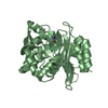
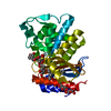
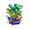
- PDB-2e6v: Crystal structure of VIP36 exoplasmic/lumenal domain, Ca2+/Man3Gl... -
+ Open data

- Basic information
| Entry | Database: PDB / ID: 2e6v | |||||||||
|---|---|---|---|---|---|---|---|---|---|---|
| Title | Crystal structure of VIP36 exoplasmic/lumenal domain, Ca2+/Man3GlcNAc-bound form | |||||||||
 Components Components | Vesicular integral-membrane protein VIP36 | |||||||||
 Keywords Keywords | PROTEIN TRANSPORT / BETA SANDWICH / CARBOHYDRATE BINDING PROTEIN / CARGO RECEPTOR | |||||||||
| Function / homology |  Function and homology information Function and homology informationCOPII-mediated vesicle transport / Cargo concentration in the ER / COPI-coated vesicle / exocytic vesicle / retrograde vesicle-mediated transport, Golgi to endoplasmic reticulum / D-mannose binding / endoplasmic reticulum-Golgi intermediate compartment / heat shock protein binding / positive regulation of phagocytosis / protein transport ...COPII-mediated vesicle transport / Cargo concentration in the ER / COPI-coated vesicle / exocytic vesicle / retrograde vesicle-mediated transport, Golgi to endoplasmic reticulum / D-mannose binding / endoplasmic reticulum-Golgi intermediate compartment / heat shock protein binding / positive regulation of phagocytosis / protein transport / cytoplasmic vesicle / Golgi membrane / perinuclear region of cytoplasm / cell surface / Golgi apparatus / : / metal ion binding / plasma membrane Similarity search - Function | |||||||||
| Biological species |  | |||||||||
| Method |  X-RAY DIFFRACTION / SYNCHROTRON / MOLECULAR REPLACEMENT / Resolution: 2.5 Å X-RAY DIFFRACTION / SYNCHROTRON / MOLECULAR REPLACEMENT / Resolution: 2.5 Å | |||||||||
 Authors Authors | Satoh, T. / Kato, R. / Wakatsuki, S. | |||||||||
 Citation Citation | Journal: J.Biol.Chem. / Year: 2007 Title: Structural basis for recognition of high mannose type glycoproteins by mammalian transport lectin VIP36 Authors: Satoh, T. / Cowieson, N.P. / Hakamata, W. / Ideo, H. / Fukushima, K. / Kurihara, M. / Kato, R. / Yamashita, K. / Wakatsuki, S. | |||||||||
| History |
| |||||||||
| Remark 650 | HELIX DETERMINATION METHOD: AUTHOR DETERMINED | |||||||||
| Remark 700 | SHEET DETERMINATION METHOD: AUTHOR DETERMINED |
- Structure visualization
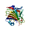
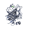
| Structure viewer | Molecule: MolmilJmol/JSmol |
|---|
- Downloads & links
-Download
| PDBx/mmCIF format | 2e6v.cif.gz | 245.4 KB | Display | PDBx/mmCIF format |
|---|---|---|---|---|
| PDB format | pdb2e6v.ent.gz | 197.8 KB | Display | PDB format |
| PDBx/mmJSON format | 2e6v.json.gz | Tree view | PDBx/mmJSON format | |
| Others |  Other downloads Other downloads |
-Validation report
| Arichive directory | https://data.pdbj.org/pub/pdb/validation_reports/e6/2e6vftp://data.pdbj.org/pub/pdb/validation_reports/e6/2e6v | HTTPS FTP |
|---|
-Related structure data
| Related structure data | 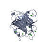 2duoC  2dupC  2duqC 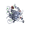 2durSC C: citing same article ( S: Starting model for refinement |
|---|---|
| Similar structure data |


-Links
 PDBj
PDBj


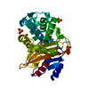
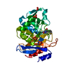
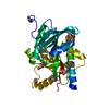
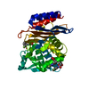
- Assembly
| Deposited unit | 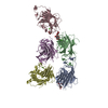
| ||||||||
|---|---|---|---|---|---|---|---|---|---|
| 1 | 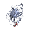
| ||||||||
| 2 | 
| ||||||||
| 3 | 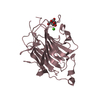
| ||||||||
| 4 | 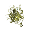
| ||||||||
| 5 | 
| ||||||||
| Unit cell |
|
-Components
| #1: Protein | Mass: 28931.318 Da / Num. of mol.: 5 / Fragment: RESIDUES 51-301 Source method: isolated from a genetically manipulated source Source: (gene. exp.)  #2: Polysaccharide | Source method: isolated from a genetically manipulated source #3: Chemical | ChemComp-CA /   Mass: 40.078 Da / Num. of mol.: 5 / Source method: obtained synthetically / Formula: Ca Mass: 40.078 Da / Num. of mol.: 5 / Source method: obtained synthetically / Formula: Ca#4: Sugar | ChemComp-MAN / |   Type: D-saccharide, alpha linking / Mass: 180.156 Da / Num. of mol.: 1 Type: D-saccharide, alpha linking / Mass: 180.156 Da / Num. of mol.: 1Source method: isolated from a genetically manipulated source Formula: C6H12O6 #5: Water | ChemComp-HOH / |  Mass: 18.015 Da / Num. of mol.: 66 / Source method: isolated from a natural source / Formula: H2O Mass: 18.015 Da / Num. of mol.: 66 / Source method: isolated from a natural source / Formula: H2OHas protein modification | Y | |
|---|
-Experimental details
-Experiment
| Experiment | Method: X-RAY DIFFRACTION / Number of used crystals: 1 |
|---|
- Sample preparation
| Crystal | Density Matthews: 2.65 Å3/Da / Density % sol: 53.52 % |
|---|---|
| Crystal grow | Temperature: 277 K / Method: vapor diffusion, hanging drop / pH: 6 Details: 10% PEG4000, 0.4M Imidazole malate (pH6.0), 3.4mM Man3GlcNAc, VAPOR DIFFUSION, HANGING DROP, temperature 277K |
-Data collection
| Diffraction | Mean temperature: 95 K |
|---|---|
| Diffraction source | Source: SYNCHROTRON / Site: Photon Factory  / Beamline: BL-5A / Wavelength: 1 Å / Beamline: BL-5A / Wavelength: 1 Å |
| Detector | Type: ADSC QUANTUM 315 / Detector: CCD / Date: Oct 24, 2006 |
| Radiation | Monochromator: Si(111) / Protocol: SINGLE WAVELENGTH / Monochromatic (M) / Laue (L): M / Scattering type: x-ray |
| Radiation wavelength | Wavelength: 1 Å / Relative weight: 1 |
| Reflection | Resolution: 2.5→50 Å / Num. all: 54228 / Num. obs: 54152 / % possible obs: 99.9 % / Redundancy: 6.5 % / Biso Wilson estimate: 44.4 Å2 / Rmerge(I) obs: 0.133 / Net I/σ(I): 8.8 |
| Reflection shell | Resolution: 2.5→2.59 Å / Redundancy: 6.6 % / Rmerge(I) obs: 0.376 / Mean I/σ(I) obs: 5.8 / Num. unique all: 5317 / % possible all: 100 |
- Processing
| Software |
| |||||||||||||||||||||||||||||||||||||||||||||||||||||||||||||||||||||||||||||||||||||||||||||||
|---|---|---|---|---|---|---|---|---|---|---|---|---|---|---|---|---|---|---|---|---|---|---|---|---|---|---|---|---|---|---|---|---|---|---|---|---|---|---|---|---|---|---|---|---|---|---|---|---|---|---|---|---|---|---|---|---|---|---|---|---|---|---|---|---|---|---|---|---|---|---|---|---|---|---|---|---|---|---|---|---|---|---|---|---|---|---|---|---|---|---|---|---|---|---|---|---|
| Refinement | Method to determine structure: MOLECULAR REPLACEMENT Starting model: PDB ENTRY 2DUR Resolution: 2.5→20 Å / Cor.coef. Fo:Fc: 0.919 / Cor.coef. Fo:Fc free: 0.876 / SU B: 9.34 / SU ML: 0.216 / Cross valid method: THROUGHOUT / σ(F): 0 / ESU R: 0.503 / ESU R Free: 0.31 / Stereochemistry target values: MAXIMUM LIKELIHOOD
| |||||||||||||||||||||||||||||||||||||||||||||||||||||||||||||||||||||||||||||||||||||||||||||||
| Solvent computation | Ion probe radii: 0.8 Å / Shrinkage radii: 0.8 Å / VDW probe radii: 1.4 Å / Solvent model: MASK | |||||||||||||||||||||||||||||||||||||||||||||||||||||||||||||||||||||||||||||||||||||||||||||||
| Displacement parameters | Biso mean: 27.96 Å2
| |||||||||||||||||||||||||||||||||||||||||||||||||||||||||||||||||||||||||||||||||||||||||||||||
| Refinement step | Cycle: LAST / Resolution: 2.5→20 Å
| |||||||||||||||||||||||||||||||||||||||||||||||||||||||||||||||||||||||||||||||||||||||||||||||
| Refine LS restraints |
| |||||||||||||||||||||||||||||||||||||||||||||||||||||||||||||||||||||||||||||||||||||||||||||||
| LS refinement shell | Highest resolution: 2.5 Å / Num. reflection Rwork: 3666 / Total num. of bins used: 20 |
