+ データを開く
- 基本情報
基本情報
| 登録情報 | データベース: PDB / ID: 7kcb | ||||||
|---|---|---|---|---|---|---|---|
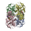
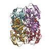
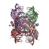
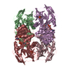
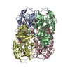
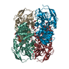
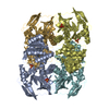
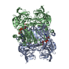
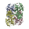
| タイトル | Symmetry in Yeast Alcohol Dehydrogenase 1 -Closed Form with NAD+ and Trifluoroethanol | ||||||
 要素 要素 | ADH1 isoform 1 | ||||||
 キーワード キーワード | OXIDOREDUCTASE / Alcohol dehydrogenase / holo-enzyme complex | ||||||
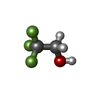
| 機能・相同性 | TRIFLUOROETHANOL / NICOTINAMIDE-ADENINE-DINUCLEOTIDE / :  機能・相同性情報 機能・相同性情報 | ||||||
| 生物種 |  | ||||||
| 手法 | 電子顕微鏡法 / 単粒子再構成法 / クライオ電子顕微鏡法 / 解像度: 2.77 Å | ||||||
 データ登録者 データ登録者 | Subramanian, R. / Chang, L. / Li, Z. / Plapp, B.V. / Guntupalli, S.R. | ||||||
 引用 引用 |  ジャーナル: Biochemistry / 年: 2021 ジャーナル: Biochemistry / 年: 2021タイトル: Cryo-Electron Microscopy Structures of Yeast Alcohol Dehydrogenase. 著者: Sai Rohit Guntupalli / Zhuang Li / Leifu Chang / Bryce V Plapp / Ramaswamy Subramanian /   要旨: Structures of yeast alcohol dehydrogenase determined by X-ray crystallography show that the subunits have two different conformational states in each of the two dimers that form the tetramer. ...Structures of yeast alcohol dehydrogenase determined by X-ray crystallography show that the subunits have two different conformational states in each of the two dimers that form the tetramer. Apoenzyme and holoenzyme complexes relevant to the catalytic mechanism were described, but the asymmetry led to questions about the cooperativity of the subunits in catalysis. This study used cryo-electron microscopy (cryo-EM) to provide structures for the apoenzyme, two different binary complexes with NADH, and a ternary complex with NAD and 2,2,2-trifluoroethanol. All four subunits in each of these complexes are identical, as the tetramers have 2 symmetry, suggesting that there is no preexisting asymmetry and that the subunits can be independently active. The apoenzyme and one enzyme-NADH complex have "open" conformations and the inverted coordination of the catalytic zinc with Cys-43, His-66, Glu-67, and Cys-153, whereas another enzyme-NADH complex and the ternary complex have closed conformations with the classical coordination of the zinc with Cys-43, His-66, Cys-153, and a water or the oxygen of trifluoroethanol. The conformational change involves interactions of Arg-340 with the pyrophosphate group of the coenzyme and Glu-67. The cryo-EM and X-ray crystallography studies provide structures relevant for the catalytic mechanism. #1:  ジャーナル: Biochemistry / 年: 2014 ジャーナル: Biochemistry / 年: 2014タイトル: Yeast alcohol dehydrogenase structure and catalysis. 著者: Savarimuthu Baskar Raj / S Ramaswamy / Bryce V Plapp /  要旨: Yeast (Saccharomyces cerevisiae) alcohol dehydrogenase I (ADH1) is the constitutive enzyme that reduces acetaldehyde to ethanol during the fermentation of glucose. ADH1 is a homotetramer of subunits ...Yeast (Saccharomyces cerevisiae) alcohol dehydrogenase I (ADH1) is the constitutive enzyme that reduces acetaldehyde to ethanol during the fermentation of glucose. ADH1 is a homotetramer of subunits with 347 amino acid residues. A structure for ADH1 was determined by X-ray crystallography at 2.4 Å resolution. The asymmetric unit contains four different subunits, arranged as similar dimers named AB and CD. The unit cell contains two different tetramers made up of "back-to-back" dimers, AB:AB and CD:CD. The A and C subunits in each dimer are structurally similar, with a closed conformation, bound coenzyme, and the oxygen of 2,2,2-trifluoroethanol ligated to the catalytic zinc in the classical tetrahedral coordination with Cys-43, Cys-153, and His-66. In contrast, the B and D subunits have an open conformation with no bound coenzyme, and the catalytic zinc has an alternative, inverted coordination with Cys-43, Cys-153, His-66, and the carboxylate of Glu-67. The asymmetry in the dimeric subunits of the tetramer provides two structures that appear to be relevant for the catalytic mechanism. The alternative coordination of the zinc may represent an intermediate in the mechanism of displacement of the zinc-bound water with alcohol or aldehyde substrates. Substitution of Glu-67 with Gln-67 decreases the catalytic efficiency by 100-fold. Previous studies of structural modeling, evolutionary relationships, substrate specificity, chemical modification, and site-directed mutagenesis are interpreted more fully with the three-dimensional structure. | ||||||
| 履歴 |
|
- 構造の表示
構造の表示
| ムービー |
 ムービービューア ムービービューア |
|---|---|
| 構造ビューア | 分子:  Molmil Molmil Jmol/JSmol Jmol/JSmol |
- ダウンロードとリンク
- ダウンロード
| PDBx/mmCIF形式 |  7kcb.cif.gz 7kcb.cif.gz | 239 KB | 表示 |  PDBx/mmCIF形式 PDBx/mmCIF形式 |
|---|---|---|---|---|
| PDB形式 |  pdb7kcb.ent.gz pdb7kcb.ent.gz | 191.5 KB | 表示 |  PDB形式 PDB形式 |
| PDBx/mmJSON形式 |  7kcb.json.gz 7kcb.json.gz | ツリー表示 |  PDBx/mmJSON形式 PDBx/mmJSON形式 | |
| その他 |  その他のダウンロード その他のダウンロード |
-検証レポート
| 文書・要旨 |  7kcb_validation.pdf.gz 7kcb_validation.pdf.gz | 1.2 MB | 表示 |  wwPDB検証レポート wwPDB検証レポート |
|---|---|---|---|---|
| 文書・詳細版 |  7kcb_full_validation.pdf.gz 7kcb_full_validation.pdf.gz | 1.2 MB | 表示 | |
| XML形式データ |  7kcb_validation.xml.gz 7kcb_validation.xml.gz | 41.7 KB | 表示 | |
| CIF形式データ |  7kcb_validation.cif.gz 7kcb_validation.cif.gz | 62.1 KB | 表示 | |
| アーカイブディレクトリ |  https://data.pdbj.org/pub/pdb/validation_reports/kc/7kcb https://data.pdbj.org/pub/pdb/validation_reports/kc/7kcb ftp://data.pdbj.org/pub/pdb/validation_reports/kc/7kcb ftp://data.pdbj.org/pub/pdb/validation_reports/kc/7kcb | HTTPS FTP |
-関連構造データ
- リンク
リンク
- 集合体
集合体
| 登録構造単位 | 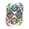
|
|---|---|
| 1 |
|
- 要素
要素
| #1: タンパク質 | 分子量: 36759.906 Da / 分子数: 4 / 由来タイプ: 天然 / 由来: (天然)  #2: 化合物 | ChemComp-ZN / #3: 化合物 | ChemComp-NAD / #4: 化合物 | ChemComp-ETF / 研究の焦点であるリガンドがあるか | Y | Has protein modification | Y | |
|---|
-実験情報
-実験
| 実験 | 手法: 電子顕微鏡法 |
|---|---|
| EM実験 | 試料の集合状態: PARTICLE / 3次元再構成法: 単粒子再構成法 |
- 試料調製
試料調製
| 構成要素 | 名称: Alcohol Dehydrogenase NAD+ Pyrazole complex / タイプ: COMPLEX / Entity ID: #1 / 由来: NATURAL |
|---|---|
| 分子量 | 値: 0.37 MDa / 実験値: NO |
| 由来(天然) | 生物種:  |
| 緩衝液 | pH: 8.2 詳細: Tris HCl buffer 5mM with 200mM KCl adjusted to pH 8.2. |
| 緩衝液成分 | 濃度: 50 mM / 名称: Tris / 式: C4H11NO3 |
| 試料 | 濃度: 5 mg/ml / 包埋: NO / シャドウイング: NO / 染色: NO / 凍結: YES / 詳細: Purified by Size Exclusion chromatography |
| 試料支持 | グリッドの材料: GOLD / グリッドのサイズ: 300 divisions/in. / グリッドのタイプ: Quantifoil |
| 急速凍結 | 装置: FEI VITROBOT MARK II / 凍結剤: ETHANE / 湿度: 100 % / 凍結前の試料温度: 298 K |
- 電子顕微鏡撮影
| 実験機器 |  モデル: Titan Krios / 画像提供: FEI Company |
|---|---|
| 顕微鏡 | モデル: FEI TITAN KRIOS |
| 電子銃 | 電子線源:  FIELD EMISSION GUN / 加速電圧: 300 kV / 照射モード: OTHER FIELD EMISSION GUN / 加速電圧: 300 kV / 照射モード: OTHER |
| 電子レンズ | モード: BRIGHT FIELD / 最大 デフォーカス(公称値): 2000 nm / 最小 デフォーカス(公称値): 800 nm / Cs: 2.7 mm / C2レンズ絞り径: 100 µm |
| 試料ホルダ | 凍結剤: NITROGEN |
| 撮影 | 電子線照射量: 54 e/Å2 フィルム・検出器のモデル: GATAN K3 BIOQUANTUM (6k x 4k) |
- 解析
解析
| EMソフトウェア |
| ||||||||||||||||||||||||
|---|---|---|---|---|---|---|---|---|---|---|---|---|---|---|---|---|---|---|---|---|---|---|---|---|---|
| CTF補正 | タイプ: NONE | ||||||||||||||||||||||||
| 3次元再構成 | 解像度: 2.77 Å / 解像度の算出法: FSC 0.143 CUT-OFF / 粒子像の数: 1284904 / アルゴリズム: BACK PROJECTION / 対称性のタイプ: POINT | ||||||||||||||||||||||||
| 原子モデル構築 | B value: 39.3 / プロトコル: RIGID BODY FIT / 空間: REAL / Target criteria: Correlation Coefficient | ||||||||||||||||||||||||
| 原子モデル構築 | PDB-ID: 5ENV PDB chain-ID: A / Accession code: 5ENV / Pdb chain residue range: 1-347 / Source name: PDB / タイプ: experimental model | ||||||||||||||||||||||||
| 精密化 | 立体化学のターゲット値: GeoStd + Monomer Library + CDL v1.2 | ||||||||||||||||||||||||
| 原子変位パラメータ | Biso mean: 43.6 Å2 | ||||||||||||||||||||||||
| 拘束条件 |
|
 ムービー
ムービー コントローラー


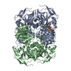
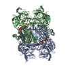

 PDBj
PDBj


