 Movie
Movie Controller

[English] 日本語
 Yorodumi
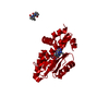
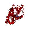
- PDB-7kaf: The crystal structure of the 2009/H1N1/California PA endonuclease... -
+ Open data

- Basic information
| Entry | Database: PDB / ID: 7kaf | ||||||
|---|---|---|---|---|---|---|---|
| Title | The crystal structure of the 2009/H1N1/California PA endonuclease I38T (construct with truncated loop 51-72) in complex with baloxavir acid | ||||||
 Components Components | Protein PA-X | ||||||
 Keywords Keywords | VIRAL PROTEIN / NUCLEASE / INFLUENZA / cleaved / DNA BINDING PROTEIN | ||||||
| Function / homology |  Function and homology information Function and homology informationsymbiont-mediated suppression of host gene expression / viral translational frameshifting / viral RNA genome replication / RNA binding / metal ion binding Similarity search - Function | ||||||
| Biological species |   Influenza A virus Influenza A virus | ||||||
| Method |  X-RAY DIFFRACTION / SYNCHROTRON / MOLECULAR REPLACEMENT / Resolution: 2.25 Å X-RAY DIFFRACTION / SYNCHROTRON / MOLECULAR REPLACEMENT / Resolution: 2.25 Å | ||||||
 Authors Authors | Cuypers, M.G. / Kumar, G. / Slavish, J. / White, S.W. | ||||||
 Citation Citation | Journal: Nucleic Acids Res. / Year: 2021 Title: Structural insights into the substrate specificity of the endonuclease activity of the influenza virus cap-snatching mechanism. Authors: Kumar, G. / Cuypers, M. / Webby, R.R. / Webb, T.R. / White, S.W. | ||||||
| History |
|
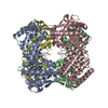
- Structure visualization
| Structure viewer | Molecule: MolmilJmol/JSmol |
|---|
- Downloads & links
-Download
| PDBx/mmCIF format | 7kaf.cif.gz | 98.7 KB | Display | PDBx/mmCIF format |
|---|---|---|---|---|
| PDB format | pdb7kaf.ent.gz | 72.7 KB | Display | PDB format |
| PDBx/mmJSON format | 7kaf.json.gz | Tree view | PDBx/mmJSON format | |
| Others |  Other downloads Other downloads |
-Validation report
| Arichive directory | https://data.pdbj.org/pub/pdb/validation_reports/ka/7kafftp://data.pdbj.org/pub/pdb/validation_reports/ka/7kaf | HTTPS FTP |
|---|
-Related structure data
| Related structure data | 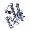 6w7aC 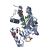 6whmC 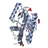 6ws3C 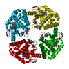 7kbcC 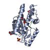 7kl3C 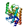 5vptS S: Starting model for refinement C: citing same article ( |
|---|---|
| Similar structure data |


-Links
 PDBj
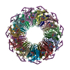
PDBj- Assembly
| Deposited unit | 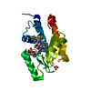
| ||||||||
|---|---|---|---|---|---|---|---|---|---|
| 1 | x 8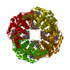
| ||||||||
| Unit cell |
|
-Components
-Protein , 1 types, 1 molecules A
| #1: Protein | Mass: 23136.289 Da / Num. of mol.: 1 / Mutation: i38T Source method: isolated from a genetically manipulated source Source: (gene. exp.) Influenza A virus, (gene. exp.) Influenza A virus (A/Luxembourg/43/2009(H1N1))Gene: PA-X, PA / Strain: A/Luxembourg/43/2009(H1N1) / Production host:  |
|---|
-Non-polymers , 5 types, 68 molecules 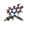


| #2: Chemical | ChemComp-E4Z /  Mass: 483.487 Da / Num. of mol.: 1 / Source method: obtained synthetically / Formula: C24H19F2N3O4S / Feature type: SUBJECT OF INVESTIGATION Mass: 483.487 Da / Num. of mol.: 1 / Source method: obtained synthetically / Formula: C24H19F2N3O4S / Feature type: SUBJECT OF INVESTIGATION | ||||
|---|---|---|---|---|---|
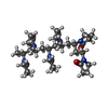
| #3: Chemical | ChemComp-QQ4 /  Mass: 668.866 Da / Num. of mol.: 1 / Source method: obtained synthetically / Formula: C36H56N6O6 Mass: 668.866 Da / Num. of mol.: 1 / Source method: obtained synthetically / Formula: C36H56N6O6 | ||||
| #4: Chemical |  Mass: 54.938 Da / Num. of mol.: 2 / Source method: obtained synthetically / Formula: Mn Mass: 54.938 Da / Num. of mol.: 2 / Source method: obtained synthetically / Formula: Mn#5: Chemical |  Mass: 96.063 Da / Num. of mol.: 2 / Source method: obtained synthetically / Formula: SO4 Mass: 96.063 Da / Num. of mol.: 2 / Source method: obtained synthetically / Formula: SO4#6: Water | ChemComp-HOH / | Mass: 18.015 Da / Num. of mol.: 62 / Source method: isolated from a natural source / Formula: H2O |
-Details
| Has ligand of interest | Y |
|---|
-Experimental details
-Experiment
| Experiment | Method: X-RAY DIFFRACTION / Number of used crystals: 1 |
|---|
- Sample preparation
| Crystal | Density Matthews: 2.92 Å3/Da / Density % sol: 57.81 % |
|---|---|
| Crystal grow | Temperature: 291 K / Method: vapor diffusion, hanging drop Details: 0.1 M HEPES PH 7.8, 1 M AMMONIUM SULFATE, 10mM MnCl2, 10mM MgCl2, 0.5% Polyvinylpyrrolidone K15 |
-Data collection
| Diffraction | Mean temperature: 100 K / Serial crystal experiment: N |
|---|---|
| Diffraction source | Source: SYNCHROTRON / Site: APS  / Beamline: 22-BM / Wavelength: 1 Å / Beamline: 22-BM / Wavelength: 1 Å |
| Detector | Type: MARMOSAIC 225 mm CCD / Detector: CCD / Date: Jun 27, 2018 |
| Radiation | Protocol: SINGLE WAVELENGTH / Monochromatic (M) / Laue (L): M / Scattering type: x-ray |
| Radiation wavelength | Wavelength: 1 Å / Relative weight: 1 |
| Reflection | Resolution: 2.25→46.06 Å / Num. obs: 12419 / % possible obs: 93.1 % / Redundancy: 9.3 % / Biso Wilson estimate: 46 Å2 / CC1/2: 1 / Rmerge(I) obs: 0.064 / Rpim(I) all: 0.032 / Rrim(I) all: 0.104 / Net I/σ(I): 12 |
| Reflection shell | Resolution: 2.25→2.33 Å / Rmerge(I) obs: 0.556 / Mean I/σ(I) obs: 1.4 / Num. unique obs: 762 / CC1/2: 0.679 / Rpim(I) all: 0.332 / Rrim(I) all: 0.744 |
- Processing
| Software |
| ||||||||||||||||||||||||||||||||||||||||||||||||||||||||||||||||||||||||||||||||||||||||||||||||||||
|---|---|---|---|---|---|---|---|---|---|---|---|---|---|---|---|---|---|---|---|---|---|---|---|---|---|---|---|---|---|---|---|---|---|---|---|---|---|---|---|---|---|---|---|---|---|---|---|---|---|---|---|---|---|---|---|---|---|---|---|---|---|---|---|---|---|---|---|---|---|---|---|---|---|---|---|---|---|---|---|---|---|---|---|---|---|---|---|---|---|---|---|---|---|---|---|---|---|---|---|---|---|
| Refinement | Method to determine structure: MOLECULAR REPLACEMENT Starting model: 5vpt Resolution: 2.25→46.06 Å / SU ML: 0.26 / Cross valid method: THROUGHOUT / σ(F): 1.36 / Phase error: 28.94 / Stereochemistry target values: ML
| ||||||||||||||||||||||||||||||||||||||||||||||||||||||||||||||||||||||||||||||||||||||||||||||||||||
| Solvent computation | Shrinkage radii: 0.9 Å / VDW probe radii: 1.11 Å / Solvent model: FLAT BULK SOLVENT MODEL | ||||||||||||||||||||||||||||||||||||||||||||||||||||||||||||||||||||||||||||||||||||||||||||||||||||
| Displacement parameters | Biso max: 142.07 Å2 / Biso mean: 60.1328 Å2 / Biso min: 32.26 Å2 | ||||||||||||||||||||||||||||||||||||||||||||||||||||||||||||||||||||||||||||||||||||||||||||||||||||
| Refinement step | Cycle: final / Resolution: 2.25→46.06 Å
| ||||||||||||||||||||||||||||||||||||||||||||||||||||||||||||||||||||||||||||||||||||||||||||||||||||
| LS refinement shell | Refine-ID: X-RAY DIFFRACTION / Rfactor Rfree error: 0 / Total num. of bins used: 5
| ||||||||||||||||||||||||||||||||||||||||||||||||||||||||||||||||||||||||||||||||||||||||||||||||||||
| Refinement TLS params. | Method: refined / Refine-ID: X-RAY DIFFRACTION
| ||||||||||||||||||||||||||||||||||||||||||||||||||||||||||||||||||||||||||||||||||||||||||||||||||||
| Refinement TLS group |
|
