 Movie
Movie Controller

[English] 日本語
 Yorodumi
- PDB-7jtt: The external aldimine crystal structure of Salmonella typhimurium... -
+ Open data

- Basic information
| Entry | Database: PDB / ID: 7jtt | ||||||
|---|---|---|---|---|---|---|---|
| Title | The external aldimine crystal structure of Salmonella typhimurium Tryptophan Synthase mutant beta-S377A in complex F9 inhibitor at the alpha-site and cesium ion at the metal coordination site. The single beta-Q114 rotamer conformation allows a hydrogen bond to form with the PLP oxygen at the position 3 in the ring | ||||||
 Components Components | (Tryptophan synthase ...) x 2 | ||||||
 Keywords Keywords | LYASE/LYASE INHIBITOR / LYASE / inhibitor complex / tryptophan synthase / LYASE-LYASE INHIBITOR complex | ||||||
| Function / homology |  Function and homology information Function and homology informationtryptophan synthase / tryptophan synthase activity / L-tryptophan biosynthetic process / identical protein binding / cytoplasm / cytosol Similarity search - Function | ||||||
| Biological species |  Salmonella typhimurium (bacteria) Salmonella typhimurium (bacteria) | ||||||
| Method |  X-RAY DIFFRACTION / MOLECULAR REPLACEMENT / molecular replacement / Resolution: 1.64 Å X-RAY DIFFRACTION / MOLECULAR REPLACEMENT / molecular replacement / Resolution: 1.64 Å | ||||||
 Authors Authors | Hilario, E. / Dunn, M.F. / Mueller, L.J. | ||||||
| Funding support |  United States, 1items United States, 1items
| ||||||
 Citation Citation | Journal: To be Published Title: The external aldimine crystal structure of Salmonella typhimurium Tryptophan Synthase mutant beta-S377A in complex F9 inhibitor at the alpha-site and cesium ion at the metal coordination site. ...Title: The external aldimine crystal structure of Salmonella typhimurium Tryptophan Synthase mutant beta-S377A in complex F9 inhibitor at the alpha-site and cesium ion at the metal coordination site. The single beta-Q114 rotamer conformation allows a hydrogen bond to form with the PLP oxygen at the position 3 in the ring. Authors: Hilario, E. / Dunn, M.F. / Mueller, L.J. | ||||||
| History |
|
- Structure visualization
| Structure viewer | Molecule: MolmilJmol/JSmol |
|---|
- Downloads & links
-Download
| PDBx/mmCIF format | 7jtt.cif.gz | 164.4 KB | Display | PDBx/mmCIF format |
|---|---|---|---|---|
| PDB format | pdb7jtt.ent.gz | 123.7 KB | Display | PDB format |
| PDBx/mmJSON format | 7jtt.json.gz | Tree view | PDBx/mmJSON format | |
| Others |  Other downloads Other downloads |
-Validation report
| Arichive directory | https://data.pdbj.org/pub/pdb/validation_reports/jt/7jttftp://data.pdbj.org/pub/pdb/validation_reports/jt/7jtt | HTTPS FTP |
|---|
-Related structure data
| Related structure data |  6sxyS S: Starting model for refinement |
|---|---|
| Similar structure data |


-Links
 PDBj
PDBj
- Assembly
| Deposited unit | 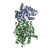
| ||||||||
|---|---|---|---|---|---|---|---|---|---|
| 1 |
| ||||||||
| Unit cell |
|
-Components
-Tryptophan synthase ... , 2 types, 2 molecules AB
| #1: Protein | Mass: 28698.797 Da / Num. of mol.: 1 Source method: isolated from a genetically manipulated source Source: (gene. exp.) Salmonella typhimurium (strain LT2 / SGSC1412 / ATCC 700720) (bacteria)Strain: LT2 / SGSC1412 / ATCC 700720 / Gene: trpA, STM1727 / Plasmid: pEBA-10 / Production host: |
|---|---|
| #2: Protein | Mass: 42902.879 Da / Num. of mol.: 1 / Mutation: S377A Source method: isolated from a genetically manipulated source Source: (gene. exp.) Salmonella typhimurium (strain LT2 / SGSC1412 / ATCC 700720) (bacteria)Strain: LT2 / SGSC1412 / ATCC 700720 / Gene: trpB, STM1726 / Plasmid: pEBA-10 / Production host: |
-Non-polymers , 8 types, 671 molecules 


| #3: Chemical | ChemComp-F9F /  Mass: 365.220 Da / Num. of mol.: 1 / Source method: obtained synthetically / Formula: C9H11F3NO7PS / Feature type: SUBJECT OF INVESTIGATION Mass: 365.220 Da / Num. of mol.: 1 / Source method: obtained synthetically / Formula: C9H11F3NO7PS / Feature type: SUBJECT OF INVESTIGATION | ||||||||||||
|---|---|---|---|---|---|---|---|---|---|---|---|---|---|
| #4: Chemical | ChemComp-DMS /  Mass: 78.133 Da / Num. of mol.: 24 / Source method: obtained synthetically / Formula: C2H6OS / Comment: DMSO, precipitant*YM Mass: 78.133 Da / Num. of mol.: 24 / Source method: obtained synthetically / Formula: C2H6OS / Comment: DMSO, precipitant*YM#5: Chemical | ChemComp-CL /  Mass: 35.453 Da / Num. of mol.: 4 / Source method: obtained synthetically / Formula: Cl Mass: 35.453 Da / Num. of mol.: 4 / Source method: obtained synthetically / Formula: Cl#6: Chemical | ChemComp-KOU / ( |  Mass: 334.219 Da / Num. of mol.: 1 / Source method: obtained synthetically / Formula: C11H15N2O8P / Feature type: SUBJECT OF INVESTIGATION Mass: 334.219 Da / Num. of mol.: 1 / Source method: obtained synthetically / Formula: C11H15N2O8P / Feature type: SUBJECT OF INVESTIGATION#7: Chemical | ChemComp-PEG / |  Mass: 106.120 Da / Num. of mol.: 1 / Source method: obtained synthetically / Formula: C4H10O3 Mass: 106.120 Da / Num. of mol.: 1 / Source method: obtained synthetically / Formula: C4H10O3#8: Chemical | ChemComp-EDO /  Mass: 62.068 Da / Num. of mol.: 7 / Source method: obtained synthetically / Formula: C2H6O2 Mass: 62.068 Da / Num. of mol.: 7 / Source method: obtained synthetically / Formula: C2H6O2#9: Chemical |  Mass: 132.905 Da / Num. of mol.: 2 / Source method: obtained synthetically / Formula: Cs / Feature type: SUBJECT OF INVESTIGATION Mass: 132.905 Da / Num. of mol.: 2 / Source method: obtained synthetically / Formula: Cs / Feature type: SUBJECT OF INVESTIGATION#10: Water | ChemComp-HOH / | Mass: 18.015 Da / Num. of mol.: 631 / Source method: isolated from a natural source / Formula: H2O |
-Details
| Has ligand of interest | Y |
|---|
-Experimental details
-Experiment
| Experiment | Method: X-RAY DIFFRACTION / Number of used crystals: 1 |
|---|
- Sample preparation
| Crystal | Density Matthews: 2.45 Å3/Da / Density % sol: 49.9 % / Description: Large plate-like crystal |
|---|---|
| Crystal grow | Temperature: 298 K / Method: vapor diffusion, sitting drop / pH: 7.8 Details: 50 mM Bicine-CsOH, 10% PEG 8,000, 4 mM Spermine, pH 7.8 PH range: 7.6-8.0 / Temp details: constant |
-Data collection
| Diffraction | Mean temperature: 100 K / Ambient temp details: Oxford Cobra System / Serial crystal experiment: N | |||||||||||||||||||||||||||||||||||||||||||||||||||||||||||||||||||||||||||||||||||||||||||||||||||
|---|---|---|---|---|---|---|---|---|---|---|---|---|---|---|---|---|---|---|---|---|---|---|---|---|---|---|---|---|---|---|---|---|---|---|---|---|---|---|---|---|---|---|---|---|---|---|---|---|---|---|---|---|---|---|---|---|---|---|---|---|---|---|---|---|---|---|---|---|---|---|---|---|---|---|---|---|---|---|---|---|---|---|---|---|---|---|---|---|---|---|---|---|---|---|---|---|---|---|---|---|
| Diffraction source | Source: ROTATING ANODE / Type: RIGAKU MICROMAX-007 HF / Wavelength: 1.5418 Å | |||||||||||||||||||||||||||||||||||||||||||||||||||||||||||||||||||||||||||||||||||||||||||||||||||
| Detector | Type: RIGAKU RAXIS IV++ / Detector: IMAGE PLATE / Date: Jan 11, 2020 / Details: Osmic Varimax HF ArcSec | |||||||||||||||||||||||||||||||||||||||||||||||||||||||||||||||||||||||||||||||||||||||||||||||||||
| Radiation | Protocol: SINGLE WAVELENGTH / Monochromatic (M) / Laue (L): M / Scattering type: x-ray | |||||||||||||||||||||||||||||||||||||||||||||||||||||||||||||||||||||||||||||||||||||||||||||||||||
| Radiation wavelength | Wavelength: 1.5418 Å / Relative weight: 1 | |||||||||||||||||||||||||||||||||||||||||||||||||||||||||||||||||||||||||||||||||||||||||||||||||||
| Reflection | Resolution: 1.64→90.091 Å / Num. all: 78381 / Num. obs: 78381 / % possible obs: 92.3 % / Redundancy: 3.4 % / Rpim(I) all: 0.053 / Rrim(I) all: 0.1 / Rsym value: 0.074 / Net I/av σ(I): 6.5 / Net I/σ(I): 7.3 / Num. measured all: 263605 | |||||||||||||||||||||||||||||||||||||||||||||||||||||||||||||||||||||||||||||||||||||||||||||||||||
| Reflection shell | Diffraction-ID: 1
|
-Phasing
| Phasing | Method: molecular replacement | ||||||||||||||||||||||||||||||||||||||||||||||||||||||||||||||||||||||||||||||||||||||||||||||||||||||||||||||||||||||||
|---|---|---|---|---|---|---|---|---|---|---|---|---|---|---|---|---|---|---|---|---|---|---|---|---|---|---|---|---|---|---|---|---|---|---|---|---|---|---|---|---|---|---|---|---|---|---|---|---|---|---|---|---|---|---|---|---|---|---|---|---|---|---|---|---|---|---|---|---|---|---|---|---|---|---|---|---|---|---|---|---|---|---|---|---|---|---|---|---|---|---|---|---|---|---|---|---|---|---|---|---|---|---|---|---|---|---|---|---|---|---|---|---|---|---|---|---|---|---|---|---|---|
| Phasing MR | R rigid body: 0.395
| ||||||||||||||||||||||||||||||||||||||||||||||||||||||||||||||||||||||||||||||||||||||||||||||||||||||||||||||||||||||||
| Phasing dm | Method: Solvent flattening and Histogram matching / Reflection: 78047 | ||||||||||||||||||||||||||||||||||||||||||||||||||||||||||||||||||||||||||||||||||||||||||||||||||||||||||||||||||||||||
| Phasing dm shell |
|
- Processing
| Software |
| |||||||||||||||||||||||||||||||||||||||||||||
|---|---|---|---|---|---|---|---|---|---|---|---|---|---|---|---|---|---|---|---|---|---|---|---|---|---|---|---|---|---|---|---|---|---|---|---|---|---|---|---|---|---|---|---|---|---|---|
| Refinement | Method to determine structure: MOLECULAR REPLACEMENT Starting model: 6SXY Resolution: 1.64→38.82 Å / Cor.coef. Fo:Fc: 0.958 / Cor.coef. Fo:Fc free: 0.943 / SU B: 2.313 / SU ML: 0.076 / SU R Cruickshank DPI: 0.1104 / Cross valid method: THROUGHOUT / σ(F): 0 / ESU R: 0.11 / ESU R Free: 0.106 / Stereochemistry target values: MAXIMUM LIKELIHOOD / Details: U VALUES : REFINED INDIVIDUALLY
| |||||||||||||||||||||||||||||||||||||||||||||
| Solvent computation | Ion probe radii: 0.8 Å / Shrinkage radii: 0.8 Å / VDW probe radii: 1.2 Å / Solvent model: MASK | |||||||||||||||||||||||||||||||||||||||||||||
| Displacement parameters | Biso max: 95.88 Å2 / Biso mean: 21.678 Å2 / Biso min: 7.32 Å2
| |||||||||||||||||||||||||||||||||||||||||||||
| Refinement step | Cycle: final / Resolution: 1.64→38.82 Å
| |||||||||||||||||||||||||||||||||||||||||||||
| Refine LS restraints |
| |||||||||||||||||||||||||||||||||||||||||||||
| LS refinement shell | Resolution: 1.64→1.683 Å / Rfactor Rfree error: 0 / Total num. of bins used: 20
|
