 Movie
Movie Controller

[English] 日本語
 Yorodumi
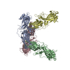
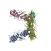
- PDB-7jtb: CRYSTAL STRUCTURE OF NATIVE BOVINE ARRESTIN 1 IN COMPLEX WITH INO... -
+ Open data

- Basic information
| Entry | Database: PDB / ID: 7jtb | ||||||
|---|---|---|---|---|---|---|---|
| Title | CRYSTAL STRUCTURE OF NATIVE BOVINE ARRESTIN 1 IN COMPLEX WITH INOSITOL HEXAKISPHOSPHATE | ||||||
 Components Components | S-arrestin | ||||||
 Keywords Keywords | PROTEIN BINDING / GPCR / rhodopsin / phototransduction / inositol / inositol hexakisphosphate / IP6 / InsP6 | ||||||
| Function / homology |  Function and homology information Function and homology informationopsin binding / Inactivation, recovery and regulation of the phototransduction cascade / G protein-coupled receptor internalization / sensory perception / response to light stimulus / photoreceptor outer segment / photoreceptor inner segment / phosphoprotein binding / G protein-coupled receptor binding / signal transduction ...opsin binding / Inactivation, recovery and regulation of the phototransduction cascade / G protein-coupled receptor internalization / sensory perception / response to light stimulus / photoreceptor outer segment / photoreceptor inner segment / phosphoprotein binding / G protein-coupled receptor binding / signal transduction / membrane / identical protein binding / cytosol Similarity search - Function | ||||||
| Biological species |  | ||||||
| Method |  X-RAY DIFFRACTION / SYNCHROTRON / FOURIER SYNTHESIS / Resolution: 2.6 Å X-RAY DIFFRACTION / SYNCHROTRON / FOURIER SYNTHESIS / Resolution: 2.6 Å | ||||||
 Authors Authors | Sander, C.L. / Palczewski, K. / Kiser, P.D. | ||||||
| Funding support |  United States, 1items United States, 1items
| ||||||
 Citation Citation | Journal: Structure / Year: 2022 Title: Structural evidence for visual arrestin priming via complexation of phosphoinositols. Authors: Sander, C.L. / Luu, J. / Kim, K. / Furkert, D. / Jang, K. / Reichenwallner, J. / Kang, M. / Lee, H.J. / Eger, B.T. / Choe, H.W. / Fiedler, D. / Ernst, O.P. / Kim, Y.J. / Palczewski, K. / Kiser, P.D. | ||||||
| History |
|
- Structure visualization
| Structure viewer | Molecule: MolmilJmol/JSmol |
|---|
- Downloads & links
-Download
| PDBx/mmCIF format | 7jtb.cif.gz | 298.4 KB | Display | PDBx/mmCIF format |
|---|---|---|---|---|
| PDB format | pdb7jtb.ent.gz | 241.8 KB | Display | PDB format |
| PDBx/mmJSON format | 7jtb.json.gz | Tree view | PDBx/mmJSON format | |
| Others |  Other downloads Other downloads |
-Validation report
| Arichive directory | https://data.pdbj.org/pub/pdb/validation_reports/jt/7jtbftp://data.pdbj.org/pub/pdb/validation_reports/jt/7jtb | HTTPS FTP |
|---|
-Related structure data
| Related structure data | 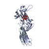 7f1wC 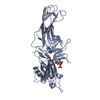 7f1xC 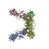 7jsmC 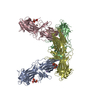 7jxaC 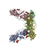 7morC 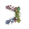 7mp0C 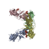 7mp1C 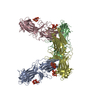 7mp2C C: citing same article ( |
|---|---|
| Similar structure data |


-Links
 PDBj
PDBj


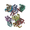
- Assembly
| Deposited unit | 
| ||||||||||||||||||||||||||||||||||||||||||||||||||||||||||||||||||||||||||||||||||||||||||||||||||||||||||||||||||||||||||||||||||||||||||||||||||||||
|---|---|---|---|---|---|---|---|---|---|---|---|---|---|---|---|---|---|---|---|---|---|---|---|---|---|---|---|---|---|---|---|---|---|---|---|---|---|---|---|---|---|---|---|---|---|---|---|---|---|---|---|---|---|---|---|---|---|---|---|---|---|---|---|---|---|---|---|---|---|---|---|---|---|---|---|---|---|---|---|---|---|---|---|---|---|---|---|---|---|---|---|---|---|---|---|---|---|---|---|---|---|---|---|---|---|---|---|---|---|---|---|---|---|---|---|---|---|---|---|---|---|---|---|---|---|---|---|---|---|---|---|---|---|---|---|---|---|---|---|---|---|---|---|---|---|---|---|---|---|---|---|
| 1 |
| ||||||||||||||||||||||||||||||||||||||||||||||||||||||||||||||||||||||||||||||||||||||||||||||||||||||||||||||||||||||||||||||||||||||||||||||||||||||
| Unit cell |
| ||||||||||||||||||||||||||||||||||||||||||||||||||||||||||||||||||||||||||||||||||||||||||||||||||||||||||||||||||||||||||||||||||||||||||||||||||||||
| Noncrystallographic symmetry (NCS) | NCS domain:
NCS domain segments: Component-ID: _ / Beg auth comp-ID: ASN / Beg label comp-ID: ASN / Refine code: _
NCS ensembles :
|
-Components
| #1: Protein | Mass: 45334.781 Da / Num. of mol.: 4 / Source method: isolated from a natural source / Source: (natural) #2: Chemical | ChemComp-IHP /   Mass: 660.035 Da / Num. of mol.: 4 / Source method: obtained synthetically / Formula: C6H18O24P6 / Feature type: SUBJECT OF INVESTIGATION Mass: 660.035 Da / Num. of mol.: 4 / Source method: obtained synthetically / Formula: C6H18O24P6 / Feature type: SUBJECT OF INVESTIGATION#3: Water | ChemComp-HOH / |  Mass: 18.015 Da / Num. of mol.: 175 / Source method: isolated from a natural source / Formula: H2O Mass: 18.015 Da / Num. of mol.: 175 / Source method: isolated from a natural source / Formula: H2OHas ligand of interest | Y | |
|---|
-Experimental details
-Experiment
| Experiment | Method: X-RAY DIFFRACTION / Number of used crystals: 1 |
|---|
- Sample preparation
| Crystal | Density Matthews: 3.92 Å3/Da / Density % sol: 68.6 % |
|---|---|
| Crystal grow | Temperature: 281 K / Method: vapor diffusion, hanging drop / pH: 7 Details: 0.1 M Bis-Tris propane, 35% 2-Ethoxyethanol, 0.001 M Magnesium acetate, 0.0009 M Phytic acid sodium salt |
-Data collection
| Diffraction | Mean temperature: 100 K / Serial crystal experiment: N |
|---|---|
| Diffraction source | Source: SYNCHROTRON / Site: APS / Beamline: 24-ID-C / Wavelength: 0.9792 Å |
| Detector | Type: DECTRIS EIGER2 X 16M / Detector: PIXEL / Date: Oct 25, 2019 |
| Radiation | Protocol: SINGLE WAVELENGTH / Monochromatic (M) / Laue (L): M / Scattering type: x-ray |
| Radiation wavelength | Wavelength: 0.9792 Å / Relative weight: 1 |
| Reflection | Resolution: 2.6→50 Å / Num. obs: 88202 / % possible obs: 99.8 % / Redundancy: 13.1 % / Biso Wilson estimate: 77.3 Å2 / CC1/2: 0.999 / Rmerge(I) obs: 0.136 / Net I/σ(I): 13.92 |
| Reflection shell | Resolution: 2.6→2.76 Å / Rmerge(I) obs: 2.233 / Num. unique obs: 14017 / CC1/2: 0.625 / % possible all: 99.4 |
- Processing
| Software |
| |||||||||||||||||||||||||||||||||||||||||||||||||||||||||||||||||
|---|---|---|---|---|---|---|---|---|---|---|---|---|---|---|---|---|---|---|---|---|---|---|---|---|---|---|---|---|---|---|---|---|---|---|---|---|---|---|---|---|---|---|---|---|---|---|---|---|---|---|---|---|---|---|---|---|---|---|---|---|---|---|---|---|---|---|
| Refinement | Method to determine structure: FOURIER SYNTHESIS / Resolution: 2.6→49.2 Å / Cor.coef. Fo:Fc: 0.959 / Cor.coef. Fo:Fc free: 0.942 / SU B: 12.208 / SU ML: 0.225 / Cross valid method: THROUGHOUT / σ(F): 0 / ESU R: 0.307 / ESU R Free: 0.236 / Stereochemistry target values: MAXIMUM LIKELIHOOD Details: HYDROGENS HAVE BEEN ADDED IN THE RIDING POSITIONS U VALUES : REFINED INDIVIDUALLY
| |||||||||||||||||||||||||||||||||||||||||||||||||||||||||||||||||
| Solvent computation | Ion probe radii: 0.8 Å / Shrinkage radii: 0.8 Å / VDW probe radii: 1 Å / Solvent model: MASK | |||||||||||||||||||||||||||||||||||||||||||||||||||||||||||||||||
| Displacement parameters | Biso max: 217.17 Å2 / Biso mean: 78.865 Å2 / Biso min: 35.21 Å2
| |||||||||||||||||||||||||||||||||||||||||||||||||||||||||||||||||
| Refinement step | Cycle: final / Resolution: 2.6→49.2 Å
| |||||||||||||||||||||||||||||||||||||||||||||||||||||||||||||||||
| Refine LS restraints |
| |||||||||||||||||||||||||||||||||||||||||||||||||||||||||||||||||
| Refine LS restraints NCS | Refine-ID: X-RAY DIFFRACTION / Type: interatomic distance / Weight position: 0.05
| |||||||||||||||||||||||||||||||||||||||||||||||||||||||||||||||||
| LS refinement shell | Resolution: 2.6→2.667 Å / Rfactor Rfree error: 0 / Total num. of bins used: 20
|
