 Movie
Movie Controller


+ Open data

- Basic information
| Entry | Database: PDB / ID: 1ayr | ||||||
|---|---|---|---|---|---|---|---|
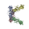
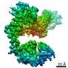
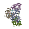
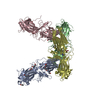
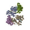
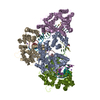
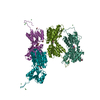
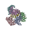
| Title | ARRESTIN FROM BOVINE ROD OUTER SEGMENTS | ||||||
 Components Components | ARRESTIN | ||||||
 Keywords Keywords | SENSORY TRANSDUCTION / ARRESTIN / RHODOPSIN | ||||||
| Function / homology |  Function and homology information Function and homology informationopsin binding / Inactivation, recovery and regulation of the phototransduction cascade / G protein-coupled receptor internalization / sensory perception / response to light stimulus / photoreceptor outer segment / photoreceptor inner segment / phosphoprotein binding / G protein-coupled receptor binding / signal transduction ...opsin binding / Inactivation, recovery and regulation of the phototransduction cascade / G protein-coupled receptor internalization / sensory perception / response to light stimulus / photoreceptor outer segment / photoreceptor inner segment / phosphoprotein binding / G protein-coupled receptor binding / signal transduction / membrane / identical protein binding / cytosol Similarity search - Function | ||||||
| Biological species |  | ||||||
| Method |  X-RAY DIFFRACTION / SYNCHROTRON / MIRAS / Resolution: 3.3 Å X-RAY DIFFRACTION / SYNCHROTRON / MIRAS / Resolution: 3.3 Å | ||||||
 Authors Authors | Granzin, J. / Wilden, U. / Choe, H.-W. / Labahn, J. / Krafft, B. / Bueldt, G. | ||||||
 Citation Citation | Journal: Nature / Year: 1998 Title: X-ray crystal structure of arrestin from bovine rod outer segments. Authors: Granzin, J. / Wilden, U. / Choe, H.W. / Labahn, J. / Krafft, B. / Buldt, G. #1: Journal: FEBS Lett. / Year: 1997Title: Crystallization and Preliminary X-Ray Analysis of Arrestin from Bovine Rod Outer Segment Authors: Wilden, U. / Choe, H.W. / Krafft, B. / Granzin, J. | ||||||
| History |
|
- Structure visualization
| Structure viewer | Molecule: MolmilJmol/JSmol |
|---|
- Downloads & links
-Download
| PDBx/mmCIF format | 1ayr.cif.gz | 272.2 KB | Display | PDBx/mmCIF format |
|---|---|---|---|---|
| PDB format | pdb1ayr.ent.gz | 225.1 KB | Display | PDB format |
| PDBx/mmJSON format | 1ayr.json.gz | Tree view | PDBx/mmJSON format | |
| Others |  Other downloads Other downloads |
-Validation report
| Arichive directory | https://data.pdbj.org/pub/pdb/validation_reports/ay/1ayrftp://data.pdbj.org/pub/pdb/validation_reports/ay/1ayr | HTTPS FTP |
|---|
-Related structure data
| Similar structure data |
|---|
-Links
 PDBj
PDBj


- Assembly
| Deposited unit | 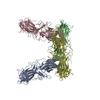
| ||||||||||||||||
|---|---|---|---|---|---|---|---|---|---|---|---|---|---|---|---|---|---|
| 1 |
| ||||||||||||||||
| Unit cell |
| ||||||||||||||||
| Noncrystallographic symmetry (NCS) | NCS oper:
|
-Components
| #1: Protein | Mass: 41001.273 Da / Num. of mol.: 4 / Source method: isolated from a natural source / Source: (natural) #2: Water | ChemComp-HOH / |  Mass: 18.015 Da / Num. of mol.: 285 / Source method: isolated from a natural source / Formula: H2O Mass: 18.015 Da / Num. of mol.: 285 / Source method: isolated from a natural source / Formula: H2OCompound details | ARRESTIN QUENCHES THE LIGHT-INDUCED ENZYME CASCADE, BINDS TO PHOSPHORYL | |
|---|
-Experimental details
-Experiment
| Experiment | Method: X-RAY DIFFRACTION / Number of used crystals: 2 |
|---|
- Sample preparation
| Crystal | Density Matthews: 3.94 Å3/Da / Density % sol: 68.5 % | ||||||||||||||||||||||||||||||||||||||||||||||||||||||||||||||||||||||||||||||||||||
|---|---|---|---|---|---|---|---|---|---|---|---|---|---|---|---|---|---|---|---|---|---|---|---|---|---|---|---|---|---|---|---|---|---|---|---|---|---|---|---|---|---|---|---|---|---|---|---|---|---|---|---|---|---|---|---|---|---|---|---|---|---|---|---|---|---|---|---|---|---|---|---|---|---|---|---|---|---|---|---|---|---|---|---|---|---|
| Crystal grow | pH: 7.2 / Details: pH 7.2 | ||||||||||||||||||||||||||||||||||||||||||||||||||||||||||||||||||||||||||||||||||||
| Crystal grow | *PLUS Method: vapor diffusion, hanging drop | ||||||||||||||||||||||||||||||||||||||||||||||||||||||||||||||||||||||||||||||||||||
| Components of the solutions | *PLUS
|
-Data collection
| Diffraction | Mean temperature: 100 K |
|---|---|
| Diffraction source | Source: SYNCHROTRON / Site: SRS  / Beamline: PX9.5 / Wavelength: 1 / Beamline: PX9.5 / Wavelength: 1 |
| Detector | Type: MARRESEARCH / Detector: IMAGE PLATE / Date: Apr 1, 1996 / Details: PT COATED TORROIDAL MIRROR |
| Radiation | Monochromator: SI(111) / Monochromatic (M) / Laue (L): M / Scattering type: x-ray |
| Radiation wavelength | Wavelength: 1 Å / Relative weight: 1 |
| Reflection | Resolution: 3.3→26.976 Å / Num. obs: 478820 / % possible obs: 95.1 % / Redundancy: 4.9 % / Rmerge(I) obs: 0.075 / Rsym value: 0.172 / Net I/σ(I): 8.6 |
| Reflection shell | Resolution: 3.3→3.39 Å / Redundancy: 2.4 % / Rmerge(I) obs: 0.172 / Mean I/σ(I) obs: 4.3 / Rsym value: 0.172 / % possible all: 79.7 |
| Reflection shell | *PLUS % possible obs: 79.7 % |
- Processing
| Software |
| ||||||||||||||||||||||||||||||||||||||||||||||||||||||||||||
|---|---|---|---|---|---|---|---|---|---|---|---|---|---|---|---|---|---|---|---|---|---|---|---|---|---|---|---|---|---|---|---|---|---|---|---|---|---|---|---|---|---|---|---|---|---|---|---|---|---|---|---|---|---|---|---|---|---|---|---|---|---|
| Refinement | Method to determine structure: MIRAS / Resolution: 3.3→130 Å / Data cutoff high absF: 1000000 / Data cutoff low absF: 0.001 / Isotropic thermal model: GROUPED, UNRESTRAINED / Cross valid method: THROUGHOUT / σ(F): 0 Details: TORSION-ANGLE MOLECULAR DYNAMICS SLOW COOL REFINEMENT
| ||||||||||||||||||||||||||||||||||||||||||||||||||||||||||||
| Displacement parameters | Biso mean: 27.71 Å2
| ||||||||||||||||||||||||||||||||||||||||||||||||||||||||||||
| Refinement step | Cycle: LAST / Resolution: 3.3→130 Å
| ||||||||||||||||||||||||||||||||||||||||||||||||||||||||||||
| Refine LS restraints |
| ||||||||||||||||||||||||||||||||||||||||||||||||||||||||||||
| Refine LS restraints NCS | NCS model details: CONSTRAINTS + RESTRAINTS | ||||||||||||||||||||||||||||||||||||||||||||||||||||||||||||
| LS refinement shell | Resolution: 3.3→3.45 Å / Total num. of bins used: 8
| ||||||||||||||||||||||||||||||||||||||||||||||||||||||||||||
| Xplor file |
| ||||||||||||||||||||||||||||||||||||||||||||||||||||||||||||
| Software | *PLUS Name: X-PLOR / Version: 3.851 / Classification: refinement | ||||||||||||||||||||||||||||||||||||||||||||||||||||||||||||
| Refinement | *PLUS | ||||||||||||||||||||||||||||||||||||||||||||||||||||||||||||
| Solvent computation | *PLUS | ||||||||||||||||||||||||||||||||||||||||||||||||||||||||||||
| Displacement parameters | *PLUS | ||||||||||||||||||||||||||||||||||||||||||||||||||||||||||||
| LS refinement shell | *PLUS Rfactor obs: 0.254 |
