 Movie
Movie Controller

[English] 日本語
 Yorodumi
- PDB-6yab: Crystal structure of PnrA from S. pneumoniae in complex with uridine -
+ Open data

- Basic information
| Entry | Database: PDB / ID: 6yab | ||||||
|---|---|---|---|---|---|---|---|
| Title | Crystal structure of PnrA from S. pneumoniae in complex with uridine | ||||||
 Components Components | (Lipoprotein) x 3 | ||||||
 Keywords Keywords | TRANSPORT PROTEIN / Nucleoside substrate-binding protein | ||||||
| Function / homology |  Function and homology information Function and homology information | ||||||
| Biological species |  Streptococcus pneumoniae serotype 4 (bacteria) Streptococcus pneumoniae serotype 4 (bacteria) | ||||||
| Method |  X-RAY DIFFRACTION / SYNCHROTRON / MOLECULAR REPLACEMENT / Resolution: 2.3 Å X-RAY DIFFRACTION / SYNCHROTRON / MOLECULAR REPLACEMENT / Resolution: 2.3 Å | ||||||
 Authors Authors | Batuecas, M.T. / Hermoso, J.A. | ||||||
| Funding support |  Spain, 1items Spain, 1items
| ||||||
 Citation Citation | Journal: J.Mol.Biol. / Year: 2021 Title: Crystal Structure and Pathophysiological Role of the Pneumococcal Nucleoside-binding Protein PnrA. Authors: Abdullah, M.R. / Batuecas, M.T. / Jennert, F. / Voss, F. / Westhoff, P. / Kohler, T.P. / Molina, R. / Hirschmann, S. / Lalk, M. / Hermoso, J.A. / Hammerschmidt, S. | ||||||
| History |
|
- Structure visualization
| Structure viewer | Molecule: MolmilJmol/JSmol |
|---|
- Downloads & links
-Download
| PDBx/mmCIF format | 6yab.cif.gz | 277.9 KB | Display | PDBx/mmCIF format |
|---|---|---|---|---|
| PDB format | pdb6yab.ent.gz | Display | PDB format | |
| PDBx/mmJSON format | 6yab.json.gz | Tree view | PDBx/mmJSON format | |
| Others |  Other downloads Other downloads |
-Validation report
| Arichive directory | https://data.pdbj.org/pub/pdb/validation_reports/ya/6yabftp://data.pdbj.org/pub/pdb/validation_reports/ya/6yab | HTTPS FTP |
|---|
-Related structure data
| Related structure data | 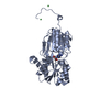 6y9uC  6ya3C  6ya4C 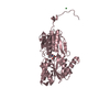 6yagC 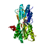 2fqwS S: Starting model for refinement C: citing same article ( |
|---|---|
| Similar structure data |


-Links
 PDBj
PDBj


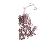
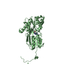
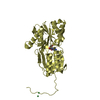
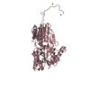
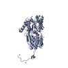
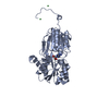
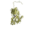
- Assembly
| Deposited unit | 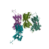
| ||||||||||||
|---|---|---|---|---|---|---|---|---|---|---|---|---|---|
| 1 | 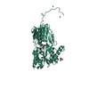
| ||||||||||||
| 2 | 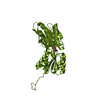
| ||||||||||||
| 3 | 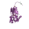
| ||||||||||||
| 4 | 
| ||||||||||||
| Unit cell |
| ||||||||||||
| Components on special symmetry positions |
|
-Components
-Protein , 3 types, 4 molecules CCCBBBAAADDD
| #1: Protein | Mass: 35127.648 Da / Num. of mol.: 2 Source method: isolated from a genetically manipulated source Source: (gene. exp.) Streptococcus pneumoniae serotype 4 (strain ATCC BAA-334 / TIGR4) (bacteria)Gene: SP_0845 / Production host: #2: Protein | | Mass: 34953.496 Da / Num. of mol.: 1 Source method: isolated from a genetically manipulated source Source: (gene. exp.) Streptococcus pneumoniae serotype 4 (strain ATCC BAA-334 / TIGR4) (bacteria)Gene: SP_0845 / Production host: #3: Protein | | Mass: 34998.469 Da / Num. of mol.: 1 Source method: isolated from a genetically manipulated source Source: (gene. exp.) Streptococcus pneumoniae serotype 4 (strain ATCC BAA-334 / TIGR4) (bacteria)Gene: SP_0845 / Production host: |
|---|
-Non-polymers , 5 types, 309 molecules 


| #4: Chemical | ChemComp-NI /  Mass: 58.693 Da / Num. of mol.: 9 / Source method: obtained synthetically / Formula: Ni Mass: 58.693 Da / Num. of mol.: 9 / Source method: obtained synthetically / Formula: Ni#5: Chemical | ChemComp-URI /  Mass: 244.201 Da / Num. of mol.: 4 / Source method: obtained synthetically / Formula: C9H12N2O6 Mass: 244.201 Da / Num. of mol.: 4 / Source method: obtained synthetically / Formula: C9H12N2O6#6: Chemical | ChemComp-CAC / |  Mass: 136.989 Da / Num. of mol.: 1 / Source method: obtained synthetically / Formula: C2H6AsO2 Mass: 136.989 Da / Num. of mol.: 1 / Source method: obtained synthetically / Formula: C2H6AsO2#7: Chemical |  Mass: 59.044 Da / Num. of mol.: 2 / Source method: obtained synthetically / Formula: C2H3O2 Mass: 59.044 Da / Num. of mol.: 2 / Source method: obtained synthetically / Formula: C2H3O2#8: Water | ChemComp-HOH / | Mass: 18.015 Da / Num. of mol.: 293 / Source method: isolated from a natural source / Formula: H2O |
|---|
-Details
| Has ligand of interest | N |
|---|
-Experimental details
-Experiment
| Experiment | Method: X-RAY DIFFRACTION / Number of used crystals: 1 |
|---|
- Sample preparation
| Crystal | Density Matthews: 2.73 Å3/Da / Density % sol: 54.86 % |
|---|---|
| Crystal grow | Temperature: 298 K / Method: vapor diffusion, hanging drop / pH: 6.5 Details: 28% PEG 600, 0.2 M calcium acetate and 0.1 M sodium cacodylate (pH 6.5) |
-Data collection
| Diffraction | Mean temperature: 100 K / Serial crystal experiment: N |
|---|---|
| Diffraction source | Source: SYNCHROTRON / Site: ALBA / Beamline: XALOC / Wavelength: 0.97895 Å |
| Detector | Type: DECTRIS PILATUS 6M / Detector: PIXEL / Date: Nov 18, 2018 |
| Radiation | Protocol: SINGLE WAVELENGTH / Monochromatic (M) / Laue (L): M / Scattering type: x-ray |
| Radiation wavelength | Wavelength: 0.97895 Å / Relative weight: 1 |
| Reflection | Resolution: 2.3→49.135 Å / Num. obs: 68314 / % possible obs: 100 % / Redundancy: 10.3 % / CC1/2: 0.99 / Rpim(I) all: 0.03 / Net I/σ(I): 17.5 |
| Reflection shell | Resolution: 2.3→2.35 Å / Mean I/σ(I) obs: 3.4 / Num. unique obs: 4545 / CC1/2: 0.93 / Rpim(I) all: 0.3 |
- Processing
| Software |
| ||||||||||||||||
|---|---|---|---|---|---|---|---|---|---|---|---|---|---|---|---|---|---|
| Refinement | Method to determine structure: MOLECULAR REPLACEMENT Starting model: 2FQW Resolution: 2.3→49.09 Å / Cross valid method: FREE R-VALUE
| ||||||||||||||||
| Refinement step | Cycle: LAST / Resolution: 2.3→49.09 Å
|
