 Movie
Movie Controller

[English] 日本語
 Yorodumi
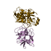
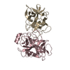
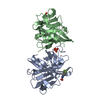
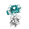
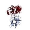
- PDB-7jrx: Crystal structure of the R64F mutant of Bauhinia Bauhinioides com... -
+ Open data

- Basic information
| Entry | Database: PDB / ID: 7jrx | ||||||
|---|---|---|---|---|---|---|---|
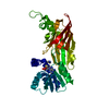
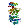
| Title | Crystal structure of the R64F mutant of Bauhinia Bauhinioides complexed with Bovine Chymotrypsin | ||||||
 Components Components |
| ||||||
 Keywords Keywords | HYDROLASE/INHIBITOR / Bovine Chymotrypsin / R64F / Bauhinia Bauhinioides Kallikrein Inhibitor / STRUCTURAL PROTEIN / HYDROLASE-INHIBITOR complex | ||||||
| Function / homology |  Function and homology information Function and homology informationchymotrypsin / endopeptidase inhibitor activity / serpin family protein binding / serine protease inhibitor complex / digestion / serine-type endopeptidase activity / proteolysis / extracellular region Similarity search - Function | ||||||
| Biological species |  Bauhinia bauhinioides (plant) Bauhinia bauhinioides (plant) | ||||||
| Method |  X-RAY DIFFRACTION / SYNCHROTRON / FOURIER SYNTHESIS / Resolution: 1.77 Å X-RAY DIFFRACTION / SYNCHROTRON / FOURIER SYNTHESIS / Resolution: 1.77 Å | ||||||
 Authors Authors | Li, M. / Wlodawer, A. / Gustchina, A. | ||||||
 Citation Citation | Journal: Acta Crystallogr D Struct Biol / Year: 2021 Title: Structural studies of complexes of kallikrein 4 with wild-type and mutated forms of the Kunitz-type inhibitor BbKI. Authors: Li, M. / Srp, J. / Mares, M. / Wlodawer, A. / Gustchina, A. | ||||||
| History |
|
- Structure visualization
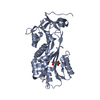
| Structure viewer | Molecule: MolmilJmol/JSmol |
|---|
- Downloads & links
-Download
| PDBx/mmCIF format | 7jrx.cif.gz | 184 KB | Display | PDBx/mmCIF format |
|---|---|---|---|---|
| PDB format | pdb7jrx.ent.gz | 143.8 KB | Display | PDB format |
| PDBx/mmJSON format | 7jrx.json.gz | Tree view | PDBx/mmJSON format | |
| Others |  Other downloads Other downloads |
-Validation report
| Arichive directory | https://data.pdbj.org/pub/pdb/validation_reports/jr/7jrxftp://data.pdbj.org/pub/pdb/validation_reports/jr/7jrx | HTTPS FTP |
|---|
-Related structure data
| Related structure data | 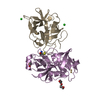 7jqkC 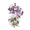 7jqnC 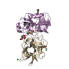 7jqoC 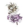 7jqvC 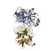 7jr1C 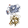 7jr2C C: citing same article ( |
|---|---|
| Similar structure data |


-Links
 PDBj
PDBj

- Assembly
| Deposited unit | 
| |||||||||||||||||||||||||||||||||||||||||||||||||||||||||||||||||||||||||||||||
|---|---|---|---|---|---|---|---|---|---|---|---|---|---|---|---|---|---|---|---|---|---|---|---|---|---|---|---|---|---|---|---|---|---|---|---|---|---|---|---|---|---|---|---|---|---|---|---|---|---|---|---|---|---|---|---|---|---|---|---|---|---|---|---|---|---|---|---|---|---|---|---|---|---|---|---|---|---|---|---|---|
| 1 | 
| |||||||||||||||||||||||||||||||||||||||||||||||||||||||||||||||||||||||||||||||
| 2 | 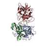
| |||||||||||||||||||||||||||||||||||||||||||||||||||||||||||||||||||||||||||||||
| Unit cell |
| |||||||||||||||||||||||||||||||||||||||||||||||||||||||||||||||||||||||||||||||
| Noncrystallographic symmetry (NCS) | NCS domain:
NCS domain segments:
NCS ensembles :
|
-Components
| #1: Protein/peptide | Mass: 1253.511 Da / Num. of mol.: 2 / Source method: isolated from a natural source / Source: (natural) #2: Protein | Mass: 13934.556 Da / Num. of mol.: 2 / Source method: isolated from a natural source / Source: (natural) #3: Protein | Mass: 10074.495 Da / Num. of mol.: 2 / Source method: isolated from a natural source / Source: (natural) #4: Protein | Mass: 18130.498 Da / Num. of mol.: 2 / Mutation: R64F Source method: isolated from a genetically manipulated source Source: (gene. exp.) Bauhinia bauhinioides (plant) / Production host:  #5: Water | ChemComp-HOH / |  Mass: 18.015 Da / Num. of mol.: 773 / Source method: isolated from a natural source / Formula: H2O Mass: 18.015 Da / Num. of mol.: 773 / Source method: isolated from a natural source / Formula: H2OHas protein modification | Y | |
|---|
-Experimental details
-Experiment
| Experiment | Method: X-RAY DIFFRACTION / Number of used crystals: 1 |
|---|
- Sample preparation
| Crystal | Density Matthews: 2.21 Å3/Da / Density % sol: 44.37 % |
|---|---|
| Crystal grow | Temperature: 293 K / Method: vapor diffusion, hanging drop / Details: 25% PEG3350, 0.2M Ammonium Acetate at pH 6.5 |
-Data collection
| Diffraction | Mean temperature: 100 K / Serial crystal experiment: N |
|---|---|
| Diffraction source | Source: SYNCHROTRON / Site: APS  / Beamline: 22-ID / Wavelength: 1 Å / Beamline: 22-ID / Wavelength: 1 Å |
| Detector | Type: DECTRIS EIGER X 16M / Detector: PIXEL / Date: Jun 19, 2020 |
| Radiation | Protocol: SINGLE WAVELENGTH / Monochromatic (M) / Laue (L): M / Scattering type: x-ray |
| Radiation wavelength | Wavelength: 1 Å / Relative weight: 1 |
| Reflection | Resolution: 1.77→85.11 Å / Num. obs: 73379 / % possible obs: 95 % / Redundancy: 6.4 % / Rsym value: 0.096 / Net I/σ(I): 12.4 |
| Reflection shell | Resolution: 1.77→1.8 Å / Num. unique obs: 2276 / Rsym value: 0.153 |
- Processing
| Software |
| ||||||||||||||||||||||||||||||||||||||||||||||||||||||||||||
|---|---|---|---|---|---|---|---|---|---|---|---|---|---|---|---|---|---|---|---|---|---|---|---|---|---|---|---|---|---|---|---|---|---|---|---|---|---|---|---|---|---|---|---|---|---|---|---|---|---|---|---|---|---|---|---|---|---|---|---|---|---|
| Refinement | Method to determine structure: FOURIER SYNTHESIS / Resolution: 1.77→71.02 Å / Cor.coef. Fo:Fc: 0.953 / Cor.coef. Fo:Fc free: 0.931 / SU B: 2.68 / SU ML: 0.083 / Cross valid method: THROUGHOUT / σ(F): 0 / ESU R: 0.125 / ESU R Free: 0.117 / Stereochemistry target values: MAXIMUM LIKELIHOOD Details: HYDROGENS HAVE BEEN ADDED IN THE RIDING POSITIONS U VALUES : REFINED INDIVIDUALLY
| ||||||||||||||||||||||||||||||||||||||||||||||||||||||||||||
| Solvent computation | Ion probe radii: 0.8 Å / Shrinkage radii: 0.8 Å / VDW probe radii: 1.2 Å / Solvent model: MASK | ||||||||||||||||||||||||||||||||||||||||||||||||||||||||||||
| Displacement parameters | Biso max: 239.55 Å2 / Biso mean: 20.979 Å2 / Biso min: 6.46 Å2
| ||||||||||||||||||||||||||||||||||||||||||||||||||||||||||||
| Refinement step | Cycle: final / Resolution: 1.77→71.02 Å
| ||||||||||||||||||||||||||||||||||||||||||||||||||||||||||||
| Refine LS restraints |
| ||||||||||||||||||||||||||||||||||||||||||||||||||||||||||||
| Refine LS restraints NCS | Refine-ID: X-RAY DIFFRACTION / Type: interatomic distance / Weight position: 0.05
| ||||||||||||||||||||||||||||||||||||||||||||||||||||||||||||
| LS refinement shell | Resolution: 1.77→1.811 Å / Rfactor Rfree error: 0
|
