 Movie
Movie Controller

[English] 日本語
 Yorodumi
- PDB-7jqn: Crystal structure of the R64M mutant of Bauhinia Bauhinioides Kal... -
+ Open data

- Basic information
| Entry | Database: PDB / ID: 7jqn | ||||||
|---|---|---|---|---|---|---|---|
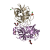
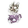
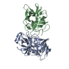
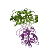
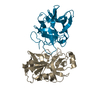
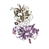
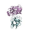
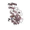
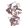
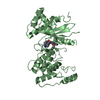
| Title | Crystal structure of the R64M mutant of Bauhinia Bauhinioides Kallikrein Inhibitor complexed with Human Kallikrein 4 | ||||||
 Components Components |
| ||||||
 Keywords Keywords | STRUCTURAL PROTEIN / HYDROLASE/INHIBITOR / Human Kallikrein 4 / Bauhinia Bauhiniordes Kallikrein Inhibitor / HYDROLASE-INHIBITOR complex | ||||||
| Function / homology |  Function and homology information Function and homology informationbiomineral tissue development / amelogenesis / endopeptidase inhibitor activity / extracellular matrix disassembly / Hydrolases; Acting on peptide bonds (peptidases); Serine endopeptidases / secretory granule / serine-type peptidase activity / protein maturation / serine-type endopeptidase activity / proteolysis ...biomineral tissue development / amelogenesis / endopeptidase inhibitor activity / extracellular matrix disassembly / Hydrolases; Acting on peptide bonds (peptidases); Serine endopeptidases / secretory granule / serine-type peptidase activity / protein maturation / serine-type endopeptidase activity / proteolysis / : / extracellular region / metal ion binding Similarity search - Function | ||||||
| Biological species |  Homo sapiens (human) Homo sapiens (human) Bauhinia bauhinioides (plant) Bauhinia bauhinioides (plant) | ||||||
| Method |  X-RAY DIFFRACTION / SYNCHROTRON / FOURIER SYNTHESIS / Resolution: 1.5 Å X-RAY DIFFRACTION / SYNCHROTRON / FOURIER SYNTHESIS / Resolution: 1.5 Å | ||||||
 Authors Authors | Li, M. / Wlodawer, A. / Gustchina, A. | ||||||
 Citation Citation | Journal: Acta Crystallogr D Struct Biol / Year: 2021 Title: Structural studies of complexes of kallikrein 4 with wild-type and mutated forms of the Kunitz-type inhibitor BbKI. Authors: Li, M. / Srp, J. / Mares, M. / Wlodawer, A. / Gustchina, A. | ||||||
| History |
|
- Structure visualization
| Structure viewer | Molecule: MolmilJmol/JSmol |
|---|
- Downloads & links
-Download
| PDBx/mmCIF format | 7jqn.cif.gz | 204.1 KB | Display | PDBx/mmCIF format |
|---|---|---|---|---|
| PDB format | pdb7jqn.ent.gz | 158.1 KB | Display | PDB format |
| PDBx/mmJSON format | 7jqn.json.gz | Tree view | PDBx/mmJSON format | |
| Others |  Other downloads Other downloads |
-Validation report
| Arichive directory | https://data.pdbj.org/pub/pdb/validation_reports/jq/7jqnftp://data.pdbj.org/pub/pdb/validation_reports/jq/7jqn | HTTPS FTP |
|---|
-Related structure data
| Related structure data | 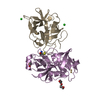 7jqkC 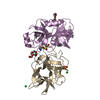 7jqoC  7jqvC 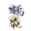 7jr1C 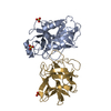 7jr2C  7jrxC C: citing same article ( |
|---|---|
| Similar structure data |


-Links
 PDBj
PDBj- Assembly
| Deposited unit | 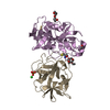
| ||||||||||||||||||
|---|---|---|---|---|---|---|---|---|---|---|---|---|---|---|---|---|---|---|---|
| 1 |
| ||||||||||||||||||
| Unit cell |
| ||||||||||||||||||
| Components on special symmetry positions |
|
-Components
-Protein , 2 types, 2 molecules EI
| #1: Protein | Mass: 23942.010 Da / Num. of mol.: 1 Source method: isolated from a genetically manipulated source Source: (gene. exp.) Homo sapiens (human) / Gene: KLK4, EMSP1, PRSS17, PSTS / Production host:  References: UniProt: Q9Y5K2, Hydrolases; Acting on peptide bonds (peptidases); Serine endopeptidases |
|---|---|
| #2: Protein | Mass: 18114.521 Da / Num. of mol.: 1 / Mutation: R64M Source method: isolated from a genetically manipulated source Source: (gene. exp.) Bauhinia bauhinioides (plant) / Production host: |
-Non-polymers , 5 types, 646 molecules 


| #3: Chemical |  Mass: 414.488 Da / Num. of mol.: 3 / Source method: obtained synthetically / Formula: C18H38O10 / Comment: precipitant*YM Mass: 414.488 Da / Num. of mol.: 3 / Source method: obtained synthetically / Formula: C18H38O10 / Comment: precipitant*YM#4: Chemical | ChemComp-CD / |  Mass: 112.411 Da / Num. of mol.: 1 / Source method: obtained synthetically / Formula: Cd Mass: 112.411 Da / Num. of mol.: 1 / Source method: obtained synthetically / Formula: Cd#5: Chemical | ChemComp-SO4 / |  Mass: 96.063 Da / Num. of mol.: 1 / Source method: obtained synthetically / Formula: SO4 Mass: 96.063 Da / Num. of mol.: 1 / Source method: obtained synthetically / Formula: SO4#6: Chemical | ChemComp-CL / |  Mass: 35.453 Da / Num. of mol.: 1 / Source method: obtained synthetically / Formula: Cl Mass: 35.453 Da / Num. of mol.: 1 / Source method: obtained synthetically / Formula: Cl#7: Water | ChemComp-HOH / | Mass: 18.015 Da / Num. of mol.: 640 / Source method: isolated from a natural source / Formula: H2O |
|---|
-Details
| Has ligand of interest | N |
|---|---|
| Has protein modification | Y |
-Experimental details
-Experiment
| Experiment | Method: X-RAY DIFFRACTION / Number of used crystals: 1 |
|---|
- Sample preparation
| Crystal | Density Matthews: 3.23 Å3/Da / Density % sol: 61.97 % |
|---|---|
| Crystal grow | Temperature: 293 K / Method: vapor diffusion, hanging drop / pH: 7.5 / Details: 2.0 Ammonium Sulfate, 2%PEG400, 10mM CdCl2 pH 7.5 |
-Data collection
| Diffraction | Mean temperature: 100 K / Serial crystal experiment: N |
|---|---|
| Diffraction source | Source: SYNCHROTRON / Site: APS  / Beamline: 22-ID / Wavelength: 1 Å / Beamline: 22-ID / Wavelength: 1 Å |
| Detector | Type: DECTRIS EIGER X 16M / Detector: PIXEL / Date: Dec 18, 2019 |
| Radiation | Protocol: SINGLE WAVELENGTH / Monochromatic (M) / Laue (L): M / Scattering type: x-ray |
| Radiation wavelength | Wavelength: 1 Å / Relative weight: 1 |
| Reflection | Resolution: 1.5→86.99 Å / Num. obs: 982025 / % possible obs: 100 % / Redundancy: 11.3 % / Rsym value: 0.083 / Net I/σ(I): 9.3 |
| Reflection shell | Resolution: 1.5→1.53 Å / Num. unique obs: 4314 / Rsym value: 0.854 |
- Processing
| Software |
| ||||||||||||||||||||||||||||||||||||||||||||||||||
|---|---|---|---|---|---|---|---|---|---|---|---|---|---|---|---|---|---|---|---|---|---|---|---|---|---|---|---|---|---|---|---|---|---|---|---|---|---|---|---|---|---|---|---|---|---|---|---|---|---|---|---|
| Refinement | Method to determine structure: FOURIER SYNTHESIS / Resolution: 1.5→62.67 Å / Cor.coef. Fo:Fc: 0.981 / Cor.coef. Fo:Fc free: 0.97 / SU B: 2.078 / SU ML: 0.034 / Cross valid method: THROUGHOUT / σ(F): 0 / ESU R: 0.05 / ESU R Free: 0.052 / Stereochemistry target values: MAXIMUM LIKELIHOOD Details: HYDROGENS HAVE BEEN USED IF PRESENT IN THE INPUT U VALUES : REFINED INDIVIDUALLY
| ||||||||||||||||||||||||||||||||||||||||||||||||||
| Solvent computation | Ion probe radii: 0.8 Å / Shrinkage radii: 0.8 Å / VDW probe radii: 1.2 Å / Solvent model: MASK | ||||||||||||||||||||||||||||||||||||||||||||||||||
| Displacement parameters | Biso max: 123.2 Å2 / Biso mean: 26.675 Å2 / Biso min: 7.81 Å2
| ||||||||||||||||||||||||||||||||||||||||||||||||||
| Refinement step | Cycle: final / Resolution: 1.5→62.67 Å
| ||||||||||||||||||||||||||||||||||||||||||||||||||
| Refine LS restraints |
| ||||||||||||||||||||||||||||||||||||||||||||||||||
| LS refinement shell | Resolution: 1.5→1.539 Å / Rfactor Rfree error: 0 / Total num. of bins used: 20
|
