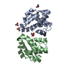
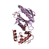
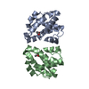
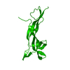
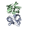
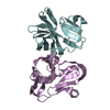
Crystal structure of EV-D68 2A protease C107A mutant
Components
Protease 2A
Keywords
VIRAL PROTEIN / HYDROLASE / metal ion binding / viral process / Enterovirus EV-D68 / protease / catalytic activity / ion binding / structural molecule activity / modulation of process of other organism / interaction with host / cellular process / gene expression / regulation of biological process / biological regulation
Function / homology
Function and homology information
picornain 2A / symbiont-mediated suppression of host mRNA export from nucleus / symbiont genome entry into host cell via pore formation in plasma membrane / picornain 3C / T=pseudo3 icosahedral viral capsid / host cell cytoplasmic vesicle membrane / ribonucleoside triphosphate phosphatase activity / nucleoside-triphosphate phosphatase / channel activity / monoatomic ion transmembrane transport ...picornain 2A / symbiont-mediated suppression of host mRNA export from nucleus / symbiont genome entry into host cell via pore formation in plasma membrane / picornain 3C / T=pseudo3 icosahedral viral capsid / host cell cytoplasmic vesicle membrane / ribonucleoside triphosphate phosphatase activity / nucleoside-triphosphate phosphatase / channel activity / monoatomic ion transmembrane transport / RNA helicase activity / symbiont-mediated suppression of host innate immune response / endocytosis involved in viral entry into host cell / symbiont-mediated activation of host autophagy / RNA-directed RNA polymerase / cysteine-type endopeptidase activity / viral RNA genome replication / RNA-directed RNA polymerase activity / virion attachment to host cell / host cell nucleus / structural molecule activity / DNA-templated transcription / proteolysis / RNA binding / zinc ion binding / ATP binding Similarity search - Function
: / Picornavirus coat protein / Poliovirus 3A protein-like / Poliovirus 3A protein like / Picornavirus 2B protein / Poliovirus core protein 3a, soluble domain / Picornavirus 2B protein / Peptidase C3, picornavirus core protein 2A / Picornavirus core protein 2A / Picornavirus coat protein VP4 ...: / Picornavirus coat protein / Poliovirus 3A protein-like / Poliovirus 3A protein like / Picornavirus 2B protein / Poliovirus core protein 3a, soluble domain / Picornavirus 2B protein / Peptidase C3, picornavirus core protein 2A / Picornavirus core protein 2A / Picornavirus coat protein VP4 / Picornavirus coat protein (VP4) / Peptidase C3A/C3B, picornaviral / 3C cysteine protease (picornain 3C) / Picornavirales 3C/3C-like protease domain / Picornavirales 3C/3C-like protease domain profile. / Picornavirus capsid / picornavirus capsid protein / Helicase, superfamily 3, single-stranded RNA virus / Superfamily 3 helicase of positive ssRNA viruses domain profile. / Helicase, superfamily 3, single-stranded DNA/RNA virus / RNA helicase / Picornavirus/Calicivirus coat protein / Viral coat protein subunit / Reverse transcriptase/Diguanylate cyclase domain / RNA-directed RNA polymerase, C-terminal domain / Viral RNA-dependent RNA polymerase / RNA-directed RNA polymerase, catalytic domain / RdRp of positive ssRNA viruses catalytic domain profile. / ATPases associated with a variety of cellular activities / AAA+ ATPase domain / Peptidase S1, PA clan, chymotrypsin-like fold / Peptidase S1, PA clan / DNA/RNA polymerase superfamily / P-loop containing nucleoside triphosphate hydrolase Similarity search - Domain/homology
In the structure databanks used in Yorodumi, some data are registered as the other names, "COVID-19 virus" and "2019-nCoV". Here are the details of the virus and the list of structure data.
Jan 31, 2019. EMDB accession codes are about to change! (news from PDBe EMDB page)
EMDB accession codes are about to change! (news from PDBe EMDB page)
The allocation of 4 digits for EMDB accession codes will soon come to an end. Whilst these codes will remain in use, new EMDB accession codes will include an additional digit and will expand incrementally as the available range of codes is exhausted. The current 4-digit format prefixed with “EMD-” (i.e. EMD-XXXX) will advance to a 5-digit format (i.e. EMD-XXXXX), and so on. It is currently estimated that the 4-digit codes will be depleted around Spring 2019, at which point the 5-digit format will come into force.
The EM Navigator/Yorodumi systems omit the EMD- prefix.
Related info.:Q: What is EMD? / ID/Accession-code notation in Yorodumi/EM Navigator
Yorodumi is a browser for structure data from EMDB, PDB, SASBDB, etc.
This page is also the successor to EM Navigator detail page, and also detail information page/front-end page for Omokage search.
The word "yorodu" (or yorozu) is an old Japanese word meaning "ten thousand". "mi" (miru) is to see.
Related info.:EMDB / PDB / SASBDB / Comparison of 3 databanks / Yorodumi Search / Aug 31, 2016. New EM Navigator & Yorodumi / Yorodumi Papers / Jmol/JSmol / Function and homology information / Changes in new EM Navigator and Yorodumi
 Movie
Movie Controller


 Open data
 Basic information
 Components
 Keywords
 Function and homology information
 Human enterovirus D68
 X-RAY DIFFRACTION /
  Authors
 United States, 1items
  Citation
 Structure visualization
 Downloads & links
 Other downloads
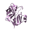


 PDBj
PDBj

 Assembly
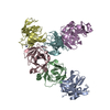


 Mass: 65.409 Da / Num. of mol.: 6 / Source method: obtained synthetically / Formula: Zn
 Mass: 18.015 Da / Num. of mol.: 59 / Source method: isolated from a natural source / Formula: H2O
 Sample preparation
 Processing