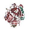
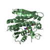
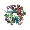
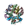
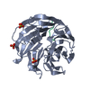
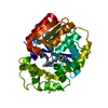
A: Insulin B chain,Insulin A chain B: Insulin B chain,Insulin A chain C: Insulin B chain,Insulin A chain D: Insulin B chain,Insulin A chain E: Insulin B chain,Insulin A chain F: Insulin B chain,Insulin A chain hetero molecules
Component-ID: 1 / Ens-ID: 1 / Beg auth comp-ID: PHE / Beg label comp-ID: PHE / End auth comp-ID: ASN / End label comp-ID: ASN / Auth seq-ID: 1 - 49 / Label seq-ID: 1 - 49
Dom-ID
Selection details
Auth asym-ID
Label asym-ID
1
chainA
A
A
2
chainB
B
B
3
chainC
C
C
4
chainD
D
D
5
chainE
E
E
6
chainF
F
F
-
Components
#1: Protein/peptide
InsulinBchain,InsulinAchain
Mass: 5601.417 Da / Num. of mol.: 6 Mutation: Residues corresponding to B chain amino acids 29-30 were deleted, and Pro-28 was mutated to Lys. This, in effect, is the equivalent of deleting only residues 28 and 30 of the wild-type B chain. Source method: obtained synthetically Details: This is a fusion protein consisting of a mutant insulin B chain followed by the insulin A chain. Six of these fusion chains are in the asymmetric unit Source: (synth.) Homo sapiens (human) / References: UniProt: P01308
Mass: 18.015 Da / Num. of mol.: 77 / Source method: isolated from a natural source / Formula: H2O
Has ligand of interest
N
Has protein modification
Y
Sequence details
This construct contains a mutant of the naturally occurring insulin B chain and the wild-type ...This construct contains a mutant of the naturally occurring insulin B chain and the wild-type naturally occurring A chain fused together into one peptide chain.
-
Experimental details
-
Experiment
Experiment
Method: X-RAY DIFFRACTION / Number of used crystals: 1
-
Sample preparation
Crystal
Density Matthews: 1.96 Å3/Da / Density % sol: 37.11 %
Crystal grow
Temperature: 298 K / Method: vapor diffusion, hanging drop Details: 1:2.5 ratio of Zn2+ to protein monomer in 0.02M Tris-HCl, 0.05M sodium citrate, 5% acetone, 0.03% phenol, 0.01% zinc acetate, PH 8.0
-
Data collection
Diffraction
Mean temperature: 100 K / Serial crystal experiment: N
In the structure databanks used in Yorodumi, some data are registered as the other names, "COVID-19 virus" and "2019-nCoV". Here are the details of the virus and the list of structure data.
Jan 31, 2019. EMDB accession codes are about to change! (news from PDBe EMDB page)
EMDB accession codes are about to change! (news from PDBe EMDB page)
The allocation of 4 digits for EMDB accession codes will soon come to an end. Whilst these codes will remain in use, new EMDB accession codes will include an additional digit and will expand incrementally as the available range of codes is exhausted. The current 4-digit format prefixed with “EMD-” (i.e. EMD-XXXX) will advance to a 5-digit format (i.e. EMD-XXXXX), and so on. It is currently estimated that the 4-digit codes will be depleted around Spring 2019, at which point the 5-digit format will come into force.
The EM Navigator/Yorodumi systems omit the EMD- prefix.
Related info.:Q: What is EMD? / ID/Accession-code notation in Yorodumi/EM Navigator
Yorodumi is a browser for structure data from EMDB, PDB, SASBDB, etc.
This page is also the successor to EM Navigator detail page, and also detail information page/front-end page for Omokage search.
The word "yorodu" (or yorozu) is an old Japanese word meaning "ten thousand". "mi" (miru) is to see.
Related info.:EMDB / PDB / SASBDB / Comparison of 3 databanks / Yorodumi Search / Aug 31, 2016. New EM Navigator & Yorodumi / Yorodumi Papers / Jmol/JSmol / Function and homology information / Changes in new EM Navigator and Yorodumi
 Movie
Movie Controller


 Open data
 Basic information
 Components
 Keywords
 Function and homology information
 Homo sapiens (human)
 X-RAY DIFFRACTION /
  Authors
 United States, 2items
  Citation
 Structure visualization
 Downloads & links
 Other downloads
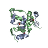


 PDBj
PDBj


 Assembly
 Mass: 94.111 Da / Num. of mol.: 6 / Source method: obtained synthetically / Formula: C6H6O
 Mass: 65.409 Da / Num. of mol.: 2 / Source method: obtained synthetically / Formula: Zn
 Mass: 35.453 Da / Num. of mol.: 2 / Source method: obtained synthetically / Formula: Cl
 Mass: 18.015 Da / Num. of mol.: 77 / Source method: isolated from a natural source / Formula: H2O
 Sample preparation
 Processing