 Movie
Movie Controller


+ Open data

- Basic information
| Entry | Database: PDB / ID: 4j82 | ||||||
|---|---|---|---|---|---|---|---|
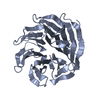
| Title | Crystal structure of beta'-COP/Insig-2 complex | ||||||
 Components Components |
| ||||||
 Keywords Keywords | PROTEIN TRANSPORT / beta propeller domain / dilysine motif / ER retrieval / vesicle trafficking | ||||||
| Function / homology |  Function and homology information Function and homology informationcranial suture morphogenesis / SREBP-SCAP-Insig complex / negative regulation of steroid biosynthetic process / SREBP-SCAP complex retention in endoplasmic reticulum / COPI vesicle coat / COPI-dependent Golgi-to-ER retrograde traffic / COPI-mediated anterograde transport / SREBP signaling pathway / late endosome to vacuole transport via multivesicular body sorting pathway / response to fatty acid ...cranial suture morphogenesis / SREBP-SCAP-Insig complex / negative regulation of steroid biosynthetic process / SREBP-SCAP complex retention in endoplasmic reticulum / COPI vesicle coat / COPI-dependent Golgi-to-ER retrograde traffic / COPI-mediated anterograde transport / SREBP signaling pathway / late endosome to vacuole transport via multivesicular body sorting pathway / response to fatty acid / negative regulation of fatty acid biosynthetic process / intracellular mRNA localization / Regulation of cholesterol biosynthesis by SREBP (SREBF) / oxysterol binding / intra-Golgi vesicle-mediated transport / retrograde vesicle-mediated transport, Golgi to endoplasmic reticulum / triglyceride metabolic process / inner ear morphogenesis / middle ear morphogenesis / cholesterol biosynthetic process / roof of mouth development / endoplasmic reticulum to Golgi vesicle-mediated transport / ubiquitin binding / protein sequestering activity / intracellular protein transport / cellular response to insulin stimulus / Golgi membrane / endoplasmic reticulum membrane / structural molecule activity / endoplasmic reticulum Similarity search - Function | ||||||
| Biological species |   Homo sapiens (human) Homo sapiens (human) | ||||||
| Method |  X-RAY DIFFRACTION / SYNCHROTRON / MOLECULAR REPLACEMENT / Resolution: 1.461 Å X-RAY DIFFRACTION / SYNCHROTRON / MOLECULAR REPLACEMENT / Resolution: 1.461 Å | ||||||
 Authors Authors | Ma, W. / Goldberg, J. | ||||||
 Citation Citation | Journal: Embo J. / Year: 2013 Title: Rules for the recognition of dilysine retrieval motifs by coatomer. Authors: Ma, W. / Goldberg, J. | ||||||
| History |
|
- Structure visualization
| Structure viewer | Molecule: MolmilJmol/JSmol |
|---|
- Downloads & links
-Download
| PDBx/mmCIF format | 4j82.cif.gz | 258.7 KB | Display | PDBx/mmCIF format |
|---|---|---|---|---|
| PDB format | pdb4j82.ent.gz | 210.1 KB | Display | PDB format |
| PDBx/mmJSON format | 4j82.json.gz | Tree view | PDBx/mmJSON format | |
| Others |  Other downloads Other downloads |
-Validation report
| Arichive directory | https://data.pdbj.org/pub/pdb/validation_reports/j8/4j82ftp://data.pdbj.org/pub/pdb/validation_reports/j8/4j82 | HTTPS FTP |
|---|
-Related structure data
| Related structure data | 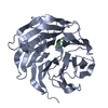 4j73C  4j77C  4j78C 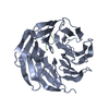 4j79C  4j81C  4j84C  4j86C  4j87C  4j8bC  4j8gC C: citing same article ( |
|---|---|
| Similar structure data |


-Links
 PDBj
PDBj

- Assembly
| Deposited unit | 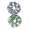
| ||||||||
|---|---|---|---|---|---|---|---|---|---|
| 1 | 
| ||||||||
| 2 | 
| ||||||||
| Unit cell |
|
-Components
| #1: Protein | Mass: 34293.652 Da / Num. of mol.: 2 / Source method: isolated from a natural source / Source: (natural) #2: Protein/peptide | Mass: 629.662 Da / Num. of mol.: 2 / Fragment: UNP residues 22-225 / Source method: obtained synthetically / Source: (synth.) Homo sapiens (human) / References: UniProt: Q9Y5U4#3: Water | ChemComp-HOH / |  Mass: 18.015 Da / Num. of mol.: 535 / Source method: isolated from a natural source / Formula: H2O Mass: 18.015 Da / Num. of mol.: 535 / Source method: isolated from a natural source / Formula: H2O |
|---|
-Experimental details
-Experiment
| Experiment | Method: X-RAY DIFFRACTION |
|---|
- Sample preparation
| Crystal | Density Matthews: 2.32 Å3/Da / Density % sol: 47.07 % |
|---|
-Data collection
| Diffraction source | Source: SYNCHROTRON / Site: APS  / Beamline: 24-ID-E / Beamline: 24-ID-E |
|---|---|
| Detector | Type: ADSC QUANTUM 315 / Detector: CCD |
| Radiation | Monochromator: Cryogenically-cooled single crystal Si(220) side bounce Protocol: SINGLE WAVELENGTH / Monochromatic (M) / Laue (L): M / Scattering type: x-ray |
| Radiation wavelength | Relative weight: 1 |
| Reflection | Resolution: 1.461→76.01 Å / Num. all: 157300 / Num. obs: 102387 / % possible obs: 95.9 % / Observed criterion σ(I): 2.2 |
- Processing
| Software | Name: PHENIX / Version: (phenix.refine: 1.7.3_928) / Classification: refinement | |||||||||||||||||||||||||||||||||||||||||||||||||||||||||||||||||||||||||||||||||||||||||||||||||||||||||
|---|---|---|---|---|---|---|---|---|---|---|---|---|---|---|---|---|---|---|---|---|---|---|---|---|---|---|---|---|---|---|---|---|---|---|---|---|---|---|---|---|---|---|---|---|---|---|---|---|---|---|---|---|---|---|---|---|---|---|---|---|---|---|---|---|---|---|---|---|---|---|---|---|---|---|---|---|---|---|---|---|---|---|---|---|---|---|---|---|---|---|---|---|---|---|---|---|---|---|---|---|---|---|---|---|---|---|
| Refinement | Method to determine structure: MOLECULAR REPLACEMENT / Resolution: 1.461→43.674 Å / SU ML: 0.22 / σ(F): 1.96 / Phase error: 25.72 / Stereochemistry target values: ML
| |||||||||||||||||||||||||||||||||||||||||||||||||||||||||||||||||||||||||||||||||||||||||||||||||||||||||
| Solvent computation | Shrinkage radii: 0.98 Å / VDW probe radii: 1.2 Å / Solvent model: FLAT BULK SOLVENT MODEL / Bsol: 55.592 Å2 / ksol: 0.389 e/Å3 | |||||||||||||||||||||||||||||||||||||||||||||||||||||||||||||||||||||||||||||||||||||||||||||||||||||||||
| Displacement parameters |
| |||||||||||||||||||||||||||||||||||||||||||||||||||||||||||||||||||||||||||||||||||||||||||||||||||||||||
| Refinement step | Cycle: LAST / Resolution: 1.461→43.674 Å
| |||||||||||||||||||||||||||||||||||||||||||||||||||||||||||||||||||||||||||||||||||||||||||||||||||||||||
| Refine LS restraints |
| |||||||||||||||||||||||||||||||||||||||||||||||||||||||||||||||||||||||||||||||||||||||||||||||||||||||||
| LS refinement shell |
|
