 Movie
Movie Controller

[English] 日本語
 Yorodumi
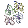
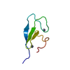
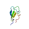
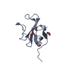
- PDB-7jku: Crystal structure of a four-tetrad, parallel, and K+ stabilized T... -
+ Open data

- Basic information
| Entry | Database: PDB / ID: 7jku | ||||||
|---|---|---|---|---|---|---|---|
| Title | Crystal structure of a four-tetrad, parallel, and K+ stabilized Tetrahymena thermophila telomeric G-quadruplex | ||||||
 Components Components | 26-mer DNA | ||||||
 Keywords Keywords | DNA / G-quadruplex / Parallel / Four-quartets / propeller loops | ||||||
| Function / homology | : / DNA / DNA (> 10) Function and homology information Function and homology information | ||||||
| Biological species |   Tetrahymena thermophila (eukaryote) Tetrahymena thermophila (eukaryote) | ||||||
| Method |  X-RAY DIFFRACTION / SYNCHROTRON / MOLECULAR REPLACEMENT / Resolution: 1.97 Å X-RAY DIFFRACTION / SYNCHROTRON / MOLECULAR REPLACEMENT / Resolution: 1.97 Å | ||||||
 Authors Authors | Yatsunyk, L.A. / McCarthy, S.E. / Beseiso, D. / Gallagher, E.P. / Chen, E.V. / Miao, J. | ||||||
| Funding support |  United States, 1items United States, 1items
| ||||||
 Citation Citation | Journal: Nucleic Acids Res. / Year: 2022 Title: The first crystal structures of hybrid and parallel four-tetrad intramolecular G-quadruplexes. Authors: Beseiso, D. / Chen, E.V. / McCarthy, S.E. / Martin, K.N. / Gallagher, E.P. / Miao, J. / Yatsunyk, L.A. | ||||||
| History |
|
- Structure visualization
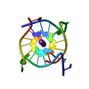
| Structure viewer | Molecule: MolmilJmol/JSmol |
|---|
- Downloads & links
-Download
| PDBx/mmCIF format | 7jku.cif.gz | 45.6 KB | Display | PDBx/mmCIF format |
|---|---|---|---|---|
| PDB format | pdb7jku.ent.gz | 27.8 KB | Display | PDB format |
| PDBx/mmJSON format | 7jku.json.gz | Tree view | PDBx/mmJSON format | |
| Others |  Other downloads Other downloads |
-Validation report
| Arichive directory | https://data.pdbj.org/pub/pdb/validation_reports/jk/7jkuftp://data.pdbj.org/pub/pdb/validation_reports/jk/7jku | HTTPS FTP |
|---|
-Related structure data


-Links
 PDBj
PDBj


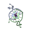
- Assembly
| Deposited unit | 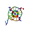
| ||||||||||||
|---|---|---|---|---|---|---|---|---|---|---|---|---|---|
| 1 |
| ||||||||||||
| Unit cell |
| ||||||||||||
| Components on special symmetry positions |
|
-Components
| #1: DNA chain | Mass: 8289.277 Da / Num. of mol.: 1 / Source method: obtained synthetically / Source: (synth.) Tetrahymena thermophila (eukaryote) | ||||
|---|---|---|---|---|---|
| #2: Chemical | ChemComp-K /   Mass: 39.098 Da / Num. of mol.: 4 / Source method: obtained synthetically / Formula: K Mass: 39.098 Da / Num. of mol.: 4 / Source method: obtained synthetically / Formula: K#3: Water | ChemComp-HOH / |  Mass: 18.015 Da / Num. of mol.: 9 / Source method: isolated from a natural source / Formula: H2O Mass: 18.015 Da / Num. of mol.: 9 / Source method: isolated from a natural source / Formula: H2OHas ligand of interest | N | |
-Experimental details
-Experiment
| Experiment | Method: X-RAY DIFFRACTION / Number of used crystals: 1 |
|---|
- Sample preparation
| Crystal | Density Matthews: 2.29 Å3/Da / Density % sol: 46.2 % |
|---|---|
| Crystal grow | Temperature: 293 K / Method: vapor diffusion, hanging drop / pH: 6.5 / Details: sodium cacodylate, KCl, PEG, NaCl, ethylene glycol |
-Data collection
| Diffraction | Mean temperature: 196 K / Serial crystal experiment: N |
|---|---|
| Diffraction source | Source: SYNCHROTRON / Site: APS / Beamline: 24-ID-E / Wavelength: 0.97918 Å |
| Detector | Type: ADSC QUANTUM 315 / Detector: CCD / Date: May 27, 2020 |
| Radiation | Protocol: SINGLE WAVELENGTH / Monochromatic (M) / Laue (L): M / Scattering type: x-ray |
| Radiation wavelength | Wavelength: 0.97918 Å / Relative weight: 1 |
| Reflection | Resolution: 1.97→59.02 Å / Num. obs: 5471 / % possible obs: 97.9 % / Redundancy: 6.3 % / Biso Wilson estimate: 56.93 Å2 / CC1/2: 0.994 / Net I/σ(I): 7.7 |
| Reflection shell | Resolution: 1.97→2.02 Å / Num. unique obs: 381 / CC1/2: 0.343 |
- Processing
| Software |
| ||||||||||||||||||||||||||||||||||||||||
|---|---|---|---|---|---|---|---|---|---|---|---|---|---|---|---|---|---|---|---|---|---|---|---|---|---|---|---|---|---|---|---|---|---|---|---|---|---|---|---|---|---|
| Refinement | Method to determine structure: MOLECULAR REPLACEMENT Starting model: TEL26-1 Resolution: 1.97→32.6 Å / SU ML: 0.34 / Cross valid method: FREE R-VALUE / σ(F): 1.35 / Phase error: 46.9033 Stereochemistry target values: GeoStd + Monomer Library + CDL v1.2
| ||||||||||||||||||||||||||||||||||||||||
| Solvent computation | Shrinkage radii: 0.9 Å / VDW probe radii: 1.11 Å / Solvent model: FLAT BULK SOLVENT MODEL | ||||||||||||||||||||||||||||||||||||||||
| Displacement parameters | Biso mean: 74.16 Å2 | ||||||||||||||||||||||||||||||||||||||||
| Refinement step | Cycle: LAST / Resolution: 1.97→32.6 Å
| ||||||||||||||||||||||||||||||||||||||||
| Refine LS restraints |
| ||||||||||||||||||||||||||||||||||||||||
| LS refinement shell |
| ||||||||||||||||||||||||||||||||||||||||
| Refinement TLS params. | Method: refined / Origin x: -4.14107878033 Å / Origin y: -16.0483557086 Å / Origin z: 6.63266892569 Å
| ||||||||||||||||||||||||||||||||||||||||
| Refinement TLS group | Selection details: all |
