 Movie
Movie Controller

[English] 日本語
 Yorodumi
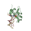
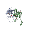
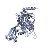
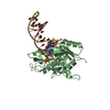
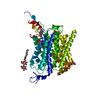
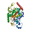
- PDB-7jk1: Human PrimPol inserting correct dCTP opposite the 8-oxoguanine lesion -
+ Open data

- Basic information
| Entry | Database: PDB / ID: 7jk1 | ||||||
|---|---|---|---|---|---|---|---|
| Title | Human PrimPol inserting correct dCTP opposite the 8-oxoguanine lesion | ||||||
 Components Components |
| ||||||
 Keywords Keywords | TRANSFERASE/DNA / Translesion / DNA synthesis / DNA Replication / DNA damage / TRANSFERASE / TRANSFERASE-DNA complex | ||||||
| Function / homology |  Function and homology information Function and homology informationDNA primase AEP / R-loop processing / mitochondrial DNA replication / DNA replication, synthesis of primer / mitochondrial DNA repair / replication fork processing / error-prone translesion synthesis / response to UV / translesion synthesis / replication fork ...DNA primase AEP / R-loop processing / mitochondrial DNA replication / DNA replication, synthesis of primer / mitochondrial DNA repair / replication fork processing / error-prone translesion synthesis / response to UV / translesion synthesis / replication fork / DNA-directed RNA polymerase complex / DNA-directed RNA polymerase activity / manganese ion binding / DNA-directed DNA polymerase / DNA-directed DNA polymerase activity / mitochondrial matrix / chromatin binding / mitochondrion / zinc ion binding / nucleus Similarity search - Function | ||||||
| Biological species |  Homo sapiens (human) Homo sapiens (human)synthetic construct (others) | ||||||
| Method |  X-RAY DIFFRACTION / SYNCHROTRON / MOLECULAR REPLACEMENT / Resolution: 2.62 Å X-RAY DIFFRACTION / SYNCHROTRON / MOLECULAR REPLACEMENT / Resolution: 2.62 Å | ||||||
 Authors Authors | Rechkoblit, O. / Aggarwal, A.K. | ||||||
| Funding support |  United States, 1items United States, 1items
| ||||||
 Citation Citation | Journal: Nat Commun / Year: 2021 Title: Structural basis of DNA synthesis opposite 8-oxoguanine by human PrimPol primase-polymerase. Authors: Rechkoblit, O. / Johnson, R.E. / Gupta, Y.K. / Prakash, L. / Prakash, S. / Aggarwal, A.K. | ||||||
| History |
|
- Structure visualization
| Structure viewer | Molecule: MolmilJmol/JSmol |
|---|
- Downloads & links
-Download
| PDBx/mmCIF format | 7jk1.cif.gz | 258.1 KB | Display | PDBx/mmCIF format |
|---|---|---|---|---|
| PDB format | pdb7jk1.ent.gz | 203.3 KB | Display | PDB format |
| PDBx/mmJSON format | 7jk1.json.gz | Tree view | PDBx/mmJSON format | |
| Others |  Other downloads Other downloads |
-Validation report
| Arichive directory | https://data.pdbj.org/pub/pdb/validation_reports/jk/7jk1ftp://data.pdbj.org/pub/pdb/validation_reports/jk/7jk1 | HTTPS FTP |
|---|
-Related structure data
| Related structure data | 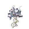 7jklC 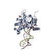 7jkpC 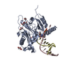 7jl8C 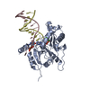 7jlgC 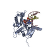 5l2xS S: Starting model for refinement C: citing same article ( |
|---|---|
| Similar structure data |


-Links
 PDBj
PDBj


- Assembly
| Deposited unit | 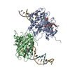
| ||||||||
|---|---|---|---|---|---|---|---|---|---|
| 1 | 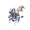
| ||||||||
| 2 | 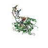
| ||||||||
| Unit cell |
|
-Components
-Protein , 1 types, 2 molecules AB
| #1: Protein | Mass: 41038.488 Da / Num. of mol.: 2 Source method: isolated from a genetically manipulated source Source: (gene. exp.) Homo sapiens (human) / Gene: PRIMPOL, CCDC111 / Production host:  References: UniProt: Q96LW4, Transferases; Transferring phosphorus-containing groups; Nucleotidyltransferases |
|---|
-DNA chain , 2 types, 4 molecules CGDF
| #2: DNA chain | Mass: 5078.291 Da / Num. of mol.: 2 / Source method: obtained synthetically / Source: (synth.) synthetic construct (others) #3: DNA chain | Mass: 3774.450 Da / Num. of mol.: 2 / Source method: obtained synthetically / Source: (synth.) synthetic construct (others) |
|---|
-Non-polymers , 5 types, 100 molecules 


| #4: Chemical |  Mass: 467.157 Da / Num. of mol.: 2 / Source method: obtained synthetically / Formula: C9H16N3O13P3 Mass: 467.157 Da / Num. of mol.: 2 / Source method: obtained synthetically / Formula: C9H16N3O13P3#5: Chemical |  Mass: 40.078 Da / Num. of mol.: 2 / Source method: obtained synthetically / Formula: Ca Mass: 40.078 Da / Num. of mol.: 2 / Source method: obtained synthetically / Formula: Ca#6: Chemical |  Mass: 92.094 Da / Num. of mol.: 3 / Source method: obtained synthetically / Formula: C3H8O3 Mass: 92.094 Da / Num. of mol.: 3 / Source method: obtained synthetically / Formula: C3H8O3#7: Chemical | ChemComp-PEG / |  Mass: 106.120 Da / Num. of mol.: 1 / Source method: obtained synthetically / Formula: C4H10O3 Mass: 106.120 Da / Num. of mol.: 1 / Source method: obtained synthetically / Formula: C4H10O3#8: Water | ChemComp-HOH / | Mass: 18.015 Da / Num. of mol.: 92 / Source method: isolated from a natural source / Formula: H2O |
|---|
-Details
| Has ligand of interest | Y |
|---|
-Experimental details
-Experiment
| Experiment | Method: X-RAY DIFFRACTION / Number of used crystals: 1 |
|---|
- Sample preparation
| Crystal | Density Matthews: 2.42 Å3/Da / Density % sol: 49.2 % |
|---|---|
| Crystal grow | Temperature: 293 K / Method: vapor diffusion, hanging drop / pH: 7.5 / Details: 225-250 mM CaCl2 16-19% PEG 3350 |
-Data collection
| Diffraction | Mean temperature: 100 K / Serial crystal experiment: N |
|---|---|
| Diffraction source | Source: SYNCHROTRON / Site: APS / Beamline: 24-ID-C / Wavelength: 0.97918 Å |
| Detector | Type: DECTRIS PILATUS3 S 6M / Detector: PIXEL / Date: Aug 16, 2016 |
| Radiation | Protocol: SINGLE WAVELENGTH / Monochromatic (M) / Laue (L): M / Scattering type: x-ray |
| Radiation wavelength | Wavelength: 0.97918 Å / Relative weight: 1 |
| Reflection | Resolution: 2.62→69.95 Å / Num. obs: 27758 / % possible obs: 98.7 % / Redundancy: 2.1 % / CC1/2: 0.988 / Rmerge(I) obs: 0.112 / Net I/σ(I): 6.1 |
| Reflection shell | Resolution: 2.62→2.74 Å / Rmerge(I) obs: 1.189 / Num. unique obs: 3360 / CC1/2: 0.375 / % possible all: 98 |
- Processing
| Software |
| ||||||||||||||||||||||||||||||||||||||||||||||||||||||||||||||||||
|---|---|---|---|---|---|---|---|---|---|---|---|---|---|---|---|---|---|---|---|---|---|---|---|---|---|---|---|---|---|---|---|---|---|---|---|---|---|---|---|---|---|---|---|---|---|---|---|---|---|---|---|---|---|---|---|---|---|---|---|---|---|---|---|---|---|---|---|
| Refinement | Method to determine structure: MOLECULAR REPLACEMENT Starting model: 5L2X Resolution: 2.62→39.877 Å / SU ML: 0.45 / Cross valid method: THROUGHOUT / σ(F): 1.96 / Phase error: 35.35 / Stereochemistry target values: ML
| ||||||||||||||||||||||||||||||||||||||||||||||||||||||||||||||||||
| Solvent computation | Shrinkage radii: 0.9 Å / VDW probe radii: 1.11 Å / Solvent model: FLAT BULK SOLVENT MODEL | ||||||||||||||||||||||||||||||||||||||||||||||||||||||||||||||||||
| Displacement parameters | Biso max: 213.3 Å2 / Biso mean: 69.95 Å2 / Biso min: 26.24 Å2 | ||||||||||||||||||||||||||||||||||||||||||||||||||||||||||||||||||
| Refinement step | Cycle: final / Resolution: 2.62→39.877 Å
| ||||||||||||||||||||||||||||||||||||||||||||||||||||||||||||||||||
| Refine LS restraints |
| ||||||||||||||||||||||||||||||||||||||||||||||||||||||||||||||||||
| LS refinement shell | Refine-ID: X-RAY DIFFRACTION / Rfactor Rfree error: 0
|
