 Movie
Movie Controller

[English] 日本語
 Yorodumi
- PDB-7jj9: Crystal structure of Zn(II)-bound AdcA from Streptococcus pneumoniae -
+ Open data

- Basic information
| Entry | Database: PDB / ID: 7jj9 | |||||||||||||||
|---|---|---|---|---|---|---|---|---|---|---|---|---|---|---|---|---|
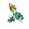
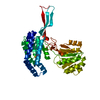
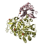
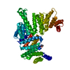
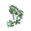
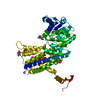
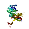
| Title | Crystal structure of Zn(II)-bound AdcA from Streptococcus pneumoniae | |||||||||||||||
 Components Components | Zinc-binding lipoprotein AdcA | |||||||||||||||
 Keywords Keywords | METAL BINDING PROTEIN / AdcA / SBP / ATP-binding cassette transporter / Zn acquisition | |||||||||||||||
| Function / homology |  Function and homology information Function and homology informationzinc ion transport / establishment of competence for transformation / cell adhesion / zinc ion binding / plasma membrane Similarity search - Function | |||||||||||||||
| Biological species |   Streptococcus pneumoniae (bacteria) Streptococcus pneumoniae (bacteria) | |||||||||||||||
| Method |  X-RAY DIFFRACTION / SYNCHROTRON / MOLECULAR REPLACEMENT / Resolution: 1.58 Å X-RAY DIFFRACTION / SYNCHROTRON / MOLECULAR REPLACEMENT / Resolution: 1.58 Å | |||||||||||||||
 Authors Authors | Luo, Z. / More, J.R. / Kobe, B. / McDevitt, C.A. | |||||||||||||||
| Funding support |  Australia, 4items Australia, 4items
| |||||||||||||||
 Citation Citation | Journal: Mbio / Year: 2021 Title: A Trap-Door Mechanism for Zinc Acquisition by Streptococcus pneumoniae AdcA. Authors: Luo, Z. / Morey, J.R. / Deplazes, E. / Motygullina, A. / Tan, A. / Ganio, K. / Neville, S.L. / Eleftheriadis, N. / Isselstein, M. / Pederick, V.G. / Paton, J.C. / Cordes, T. / Harmer, J.R. / ...Authors: Luo, Z. / Morey, J.R. / Deplazes, E. / Motygullina, A. / Tan, A. / Ganio, K. / Neville, S.L. / Eleftheriadis, N. / Isselstein, M. / Pederick, V.G. / Paton, J.C. / Cordes, T. / Harmer, J.R. / Kobe, B. / McDevitt, C.A. | |||||||||||||||
| History |
|
- Structure visualization
| Structure viewer | Molecule: MolmilJmol/JSmol |
|---|
- Downloads & links
-Download
| PDBx/mmCIF format | 7jj9.cif.gz | 199.9 KB | Display | PDBx/mmCIF format |
|---|---|---|---|---|
| PDB format | pdb7jj9.ent.gz | 156.1 KB | Display | PDB format |
| PDBx/mmJSON format | 7jj9.json.gz | Tree view | PDBx/mmJSON format | |
| Others |  Other downloads Other downloads |
-Validation report
| Arichive directory | https://data.pdbj.org/pub/pdb/validation_reports/jj/7jj9ftp://data.pdbj.org/pub/pdb/validation_reports/jj/7jj9 | HTTPS FTP |
|---|
-Related structure data
| Related structure data | 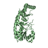 7jj8C  7jjaC  7jjbC  2osvS S: Starting model for refinement C: citing same article ( |
|---|---|
| Similar structure data |


-Links
 PDBj
PDBj


- Assembly
| Deposited unit | 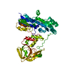
| ||||||||||||
|---|---|---|---|---|---|---|---|---|---|---|---|---|---|
| 1 |
| ||||||||||||
| Unit cell |
|
-Components
| #1: Protein | Mass: 53545.367 Da / Num. of mol.: 1 Source method: isolated from a genetically manipulated source Source: (gene. exp.) Streptococcus pneumoniae (strain ATCC BAA-255 / R6) (bacteria)Strain: ATCC BAA-255 / R6 / Gene: adcA, spr1975 / Production host: | ||||||
|---|---|---|---|---|---|---|---|
| #2: Chemical |   Mass: 65.409 Da / Num. of mol.: 2 / Source method: obtained synthetically / Formula: Zn / Feature type: SUBJECT OF INVESTIGATION Mass: 65.409 Da / Num. of mol.: 2 / Source method: obtained synthetically / Formula: Zn / Feature type: SUBJECT OF INVESTIGATION#3: Chemical | ChemComp-CL / |   Mass: 35.453 Da / Num. of mol.: 1 / Source method: obtained synthetically / Formula: Cl Mass: 35.453 Da / Num. of mol.: 1 / Source method: obtained synthetically / Formula: Cl#4: Water | ChemComp-HOH / |  Mass: 18.015 Da / Num. of mol.: 645 / Source method: isolated from a natural source / Formula: H2O Mass: 18.015 Da / Num. of mol.: 645 / Source method: isolated from a natural source / Formula: H2OHas ligand of interest | Y | |
-Experimental details
-Experiment
| Experiment | Method: X-RAY DIFFRACTION / Number of used crystals: 1 |
|---|
- Sample preparation
| Crystal | Density Matthews: 2.21 Å3/Da / Density % sol: 44.29 % |
|---|---|
| Crystal grow | Temperature: 291 K / Method: vapor diffusion, hanging drop / pH: 6.5 Details: 10% w/v polyethylene glycol 20000, 18% v/v PEG monomethyl ether 550, 0.03 M CaCl2, 0.03 M MgCl2, 0.1 M MES/imidazole pH 6.5 |
-Data collection
| Diffraction | Mean temperature: 100 K / Serial crystal experiment: N |
|---|---|
| Diffraction source | Source: SYNCHROTRON / Site: Australian Synchrotron / Beamline: MX1 / Wavelength: 0.954 Å |
| Detector | Type: ADSC QUANTUM 210r / Detector: CCD / Date: Jul 10, 2014 |
| Radiation | Protocol: SINGLE WAVELENGTH / Monochromatic (M) / Laue (L): M / Scattering type: x-ray |
| Radiation wavelength | Wavelength: 0.954 Å / Relative weight: 1 |
| Reflection | Resolution: 1.58→19.84 Å / Num. obs: 64356 / % possible obs: 99.9 % / Redundancy: 7.4 % / Biso Wilson estimate: 13.8320172758 Å2 / CC1/2: 0.99 / Rmerge(I) obs: 0.096 / Net I/σ(I): 15.9 |
| Reflection shell | Resolution: 1.58→1.61 Å / Rmerge(I) obs: 0.843 / Num. unique obs: 275 / CC1/2: 0.8 |
- Processing
| Software |
| ||||||||||||||||||||||||||||||||||||||||||||||||||||||||||||||||||||||||||||||||||||||||||||||||||||||||||||||||||||||||||||||||||||||||||||||||||||||||||||||||||||||||
|---|---|---|---|---|---|---|---|---|---|---|---|---|---|---|---|---|---|---|---|---|---|---|---|---|---|---|---|---|---|---|---|---|---|---|---|---|---|---|---|---|---|---|---|---|---|---|---|---|---|---|---|---|---|---|---|---|---|---|---|---|---|---|---|---|---|---|---|---|---|---|---|---|---|---|---|---|---|---|---|---|---|---|---|---|---|---|---|---|---|---|---|---|---|---|---|---|---|---|---|---|---|---|---|---|---|---|---|---|---|---|---|---|---|---|---|---|---|---|---|---|---|---|---|---|---|---|---|---|---|---|---|---|---|---|---|---|---|---|---|---|---|---|---|---|---|---|---|---|---|---|---|---|---|---|---|---|---|---|---|---|---|---|---|---|---|---|---|---|---|
| Refinement | Method to determine structure: MOLECULAR REPLACEMENT Starting model: 2OSV Resolution: 1.58→19.83 Å / SU ML: 0.15851032789 / Cross valid method: FREE R-VALUE / σ(F): 1.35 / Phase error: 19.4721589407 Stereochemistry target values: GeoStd + Monomer Library + CDL v1.2
| ||||||||||||||||||||||||||||||||||||||||||||||||||||||||||||||||||||||||||||||||||||||||||||||||||||||||||||||||||||||||||||||||||||||||||||||||||||||||||||||||||||||||
| Solvent computation | Shrinkage radii: 0.9 Å / VDW probe radii: 1.11 Å / Solvent model: FLAT BULK SOLVENT MODEL | ||||||||||||||||||||||||||||||||||||||||||||||||||||||||||||||||||||||||||||||||||||||||||||||||||||||||||||||||||||||||||||||||||||||||||||||||||||||||||||||||||||||||
| Displacement parameters | Biso mean: 21.7 Å2 | ||||||||||||||||||||||||||||||||||||||||||||||||||||||||||||||||||||||||||||||||||||||||||||||||||||||||||||||||||||||||||||||||||||||||||||||||||||||||||||||||||||||||
| Refinement step | Cycle: LAST / Resolution: 1.58→19.83 Å
| ||||||||||||||||||||||||||||||||||||||||||||||||||||||||||||||||||||||||||||||||||||||||||||||||||||||||||||||||||||||||||||||||||||||||||||||||||||||||||||||||||||||||
| Refine LS restraints |
| ||||||||||||||||||||||||||||||||||||||||||||||||||||||||||||||||||||||||||||||||||||||||||||||||||||||||||||||||||||||||||||||||||||||||||||||||||||||||||||||||||||||||
| LS refinement shell |
|
