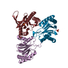
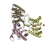
BIOMOLECULE: 1 THIS ENTRY CONTAINS THE CRYSTALLOGRAPHIC ASYMMETRIC UNIT WHICH CONSISTS OF 1 CHAIN(S) ...BIOMOLECULE: 1 THIS ENTRY CONTAINS THE CRYSTALLOGRAPHIC ASYMMETRIC UNIT WHICH CONSISTS OF 1 CHAIN(S). SEE REMARK 350 FOR INFORMATION ON GENERATING THE BIOLOGICAL MOLECULE(S). THE BIOLOGICAL UNIT IS A TETRAMER WITH 222 POINT SYMMETRY, FORMED BY CRYSTALLOGRAPHIC SYMMETRY, AS ADJUDGED BY EXTENSIVE HYDROPHOBIC CONTACTS BETWEEN THESE UNITS.
Resolution: 2→50 Å / Num. obs: 34597 / % possible obs: 100 % / Redundancy: 8.55 % / Rsym value: 0.087 / Net I/σ(I): 20.46
Reflection shell
Resolution: 2→2.07 Å / Redundancy: 7.44 % / Mean I/σ(I) obs: 3.33 / Rsym value: 0.607 / % possible all: 99.5
Reflection
*PLUS
Highest resolution: 2 Å / Lowest resolution: 50 Å / % possible obs: 99.3 % / Num. measured all: 295973 / Rmerge(I) obs: 0.087
Reflection shell
*PLUS
% possible obs: 99.5 % / Rmerge(I) obs: 0.6 / Mean I/σ(I) obs: 3.3
-
Processing
Software
Name
Version
Classification
MOSFLM
datareduction
CCP4
datareduction
SCALEPACK
datascaling
SOLVE
phasing
RESOLVE
modelbuilding
REFMAC
5.1.24
refinement
CCP4
datascaling
RESOLVE
phasing
Refinement
Method to determine structure: MAD / Resolution: 2→38.22 Å / Cor.coef. Fo:Fc: 0.96 / Cor.coef. Fo:Fc free: 0.94 / SU B: 3.584 / SU ML: 0.098 / TLS residual ADP flag: LIKELY RESIDUAL / Isotropic thermal model: Isotropic / Cross valid method: THROUGHOUT / σ(F): 0 / ESU R: 0.153 / ESU R Free: 0.143 / Stereochemistry target values: MAXIMUM LIKELIHOOD Details: THERE IS PROMINENT DIFFERENCE DENSITY ATTACHED TO CYS255, POSSIBLY THE RESULT OF PARTIAL OXIDATION, WHICH WAS HOWEVER NOT MODELLED. CNS/O WERE ALSO USED IN REFINEMENT.
Rfactor
Num. reflection
% reflection
Selection details
Rfree
0.203
2521
7.6 %
RANDOM
Rwork
0.16
-
-
-
obs
0.163
30837
95.9 %
-
Solvent computation
Ion probe radii: 0.8 Å / Shrinkage radii: 0.8 Å / VDW probe radii: 1.4 Å / Solvent model: BABINET MODEL WITH MASK
Displacement parameters
Biso mean: 18.69 Å2
Baniso -1
Baniso -2
Baniso -3
1-
-0.39 Å2
0 Å2
0 Å2
2-
-
1.02 Å2
0 Å2
3-
-
-
-0.64 Å2
Refinement step
Cycle: LAST / Resolution: 2→38.22 Å
Protein
Nucleic acid
Ligand
Solvent
Total
Num. atoms
3227
0
0
299
3526
Refine LS restraints
Refine-ID
Type
Dev ideal
Dev ideal target
Number
X-RAY DIFFRACTION
r_bond_refined_d
0.016
0.021
3284
X-RAY DIFFRACTION
r_bond_other_d
0.002
0.02
3136
X-RAY DIFFRACTION
r_angle_refined_deg
1.532
1.968
4440
X-RAY DIFFRACTION
r_angle_other_deg
0.843
3
7269
X-RAY DIFFRACTION
r_dihedral_angle_1_deg
6.116
5
413
X-RAY DIFFRACTION
r_dihedral_angle_2_deg
X-RAY DIFFRACTION
r_dihedral_angle_3_deg
X-RAY DIFFRACTION
r_dihedral_angle_4_deg
X-RAY DIFFRACTION
r_chiral_restr
0.093
0.2
524
X-RAY DIFFRACTION
r_gen_planes_refined
0.007
0.02
3618
X-RAY DIFFRACTION
r_gen_planes_other
0.006
0.02
638
X-RAY DIFFRACTION
r_nbd_refined
0.202
0.2
663
X-RAY DIFFRACTION
r_nbd_other
0.244
0.2
3606
X-RAY DIFFRACTION
r_nbtor_refined
X-RAY DIFFRACTION
r_nbtor_other
0.087
0.2
2063
X-RAY DIFFRACTION
r_xyhbond_nbd_refined
0.178
0.2
229
X-RAY DIFFRACTION
r_xyhbond_nbd_other
X-RAY DIFFRACTION
r_metal_ion_refined
X-RAY DIFFRACTION
r_metal_ion_other
X-RAY DIFFRACTION
r_symmetry_vdw_refined
0.145
0.2
20
X-RAY DIFFRACTION
r_symmetry_vdw_other
0.357
0.2
128
X-RAY DIFFRACTION
r_symmetry_hbond_refined
0.337
0.2
23
X-RAY DIFFRACTION
r_symmetry_hbond_other
X-RAY DIFFRACTION
r_mcbond_it
0.816
1.5
2059
X-RAY DIFFRACTION
r_mcangle_it
1.49
2
3342
X-RAY DIFFRACTION
r_scbond_it
2.55
3
1225
X-RAY DIFFRACTION
r_scangle_it
4.321
4.5
1098
X-RAY DIFFRACTION
r_rigid_bond_restr
X-RAY DIFFRACTION
r_sphericity_free
X-RAY DIFFRACTION
r_sphericity_bonded
LS refinement shell
Resolution: 2→2.05 Å / Total num. of bins used: 20 /
Rfactor
Num. reflection
Rfree
0.269
137
Rwork
0.196
1866
Refinement TLS params.
Method: refined / Origin x: 7.6053 Å / Origin y: 41.9533 Å / Origin z: 67.2913 Å
In the structure databanks used in Yorodumi, some data are registered as the other names, "COVID-19 virus" and "2019-nCoV". Here are the details of the virus and the list of structure data.
Jan 31, 2019. EMDB accession codes are about to change! (news from PDBe EMDB page)
EMDB accession codes are about to change! (news from PDBe EMDB page)
The allocation of 4 digits for EMDB accession codes will soon come to an end. Whilst these codes will remain in use, new EMDB accession codes will include an additional digit and will expand incrementally as the available range of codes is exhausted. The current 4-digit format prefixed with “EMD-” (i.e. EMD-XXXX) will advance to a 5-digit format (i.e. EMD-XXXXX), and so on. It is currently estimated that the 4-digit codes will be depleted around Spring 2019, at which point the 5-digit format will come into force.
The EM Navigator/Yorodumi systems omit the EMD- prefix.
Related info.:Q: What is EMD? / ID/Accession-code notation in Yorodumi/EM Navigator
Yorodumi is a browser for structure data from EMDB, PDB, SASBDB, etc.
This page is also the successor to EM Navigator detail page, and also detail information page/front-end page for Omokage search.
The word "yorodu" (or yorozu) is an old Japanese word meaning "ten thousand". "mi" (miru) is to see.
Related info.:EMDB / PDB / SASBDB / Comparison of 3 databanks / Yorodumi Search / Aug 31, 2016. New EM Navigator & Yorodumi / Yorodumi Papers / Jmol/JSmol / Function and homology information / Changes in new EM Navigator and Yorodumi
 Movie
Movie Controller

 Yorodumi
 Open data
 Basic information
 Components
 Keywords
 Function and homology information
 Thermotoga maritima (bacteria)
 X-RAY DIFFRACTION /
  Authors
 Citation
 Structure visualization
 Downloads & links
 Other downloads


 PDBj
PDBj

 Assembly
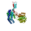

 Mass: 18.015 Da / Num. of mol.: 299 / Source method: isolated from a natural source / Formula: H2O
 Sample preparation
 / Beamline: 5.0.2 / Wavelength: 0.9793, 0.9566, 0.9794
 Processing