 Movie
Movie Controller

[English] 日本語
 Yorodumi
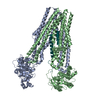
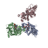
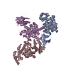
- PDB-7jix: Murine antibody that engages the influenza hemagglutinin receptor... -
+ Open data

- Basic information
| Entry | Database: PDB / ID: 7jix | ||||||
|---|---|---|---|---|---|---|---|
| Title | Murine antibody that engages the influenza hemagglutinin receptor binding site | ||||||
 Components Components |
| ||||||
 Keywords Keywords | Viral protein/IMMUNE SYSTEM / antibody / influenza / hemagglutinin / IMMUNE SYSTEM / Viral protein-IMMUNE SYSTEM complex | ||||||
| Function / homology |  Function and homology information Function and homology informationviral budding from plasma membrane / clathrin-dependent endocytosis of virus by host cell / apical plasma membrane / host cell surface receptor binding / fusion of virus membrane with host plasma membrane / fusion of virus membrane with host endosome membrane / viral envelope / virion attachment to host cell / host cell plasma membrane / virion membrane Similarity search - Function | ||||||
| Biological species |   Influenza A virus Influenza A virus | ||||||
| Method |  X-RAY DIFFRACTION / SYNCHROTRON / MOLECULAR REPLACEMENT / molecular replacement / Resolution: 3.901 Å X-RAY DIFFRACTION / SYNCHROTRON / MOLECULAR REPLACEMENT / molecular replacement / Resolution: 3.901 Å | ||||||
 Authors Authors | Bajic, G. / Schmidt, A.G. | ||||||
| Funding support |  United States, 1items United States, 1items
| ||||||
 Citation Citation | Journal: To Be Published Title: Murine antibody that engages the influenza hemagglutinin receptor binding site Authors: Bajic, G. / Schmidt, A.G. | ||||||
| History |
|
- Structure visualization
| Structure viewer | Molecule: MolmilJmol/JSmol |
|---|
- Downloads & links
-Download
| PDBx/mmCIF format | 7jix.cif.gz | 201.2 KB | Display | PDBx/mmCIF format |
|---|---|---|---|---|
| PDB format | pdb7jix.ent.gz | 156.5 KB | Display | PDB format |
| PDBx/mmJSON format | 7jix.json.gz | Tree view | PDBx/mmJSON format | |
| Others |  Other downloads Other downloads |
-Validation report
| Arichive directory | https://data.pdbj.org/pub/pdb/validation_reports/ji/7jixftp://data.pdbj.org/pub/pdb/validation_reports/ji/7jix | HTTPS FTP |
|---|
-Related structure data
| Related structure data |  5ugyS S: Starting model for refinement |
|---|---|
| Similar structure data |


-Links
 PDBj
PDBj


- Assembly
| Deposited unit | 
| ||||||||
|---|---|---|---|---|---|---|---|---|---|
| 1 | 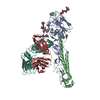
| ||||||||
| Unit cell |
|
-Components
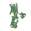
-Hemagglutinin ... , 2 types, 2 molecules AB
| #1: Protein | Mass: 37239.723 Da / Num. of mol.: 1 Source method: isolated from a genetically manipulated source Source: (gene. exp.) Influenza A virus (A/Solomon Islands/3/2006 (Egg passage)(H1N1))Strain: A/Solomon Islands/3/2006 (Egg passage)(H1N1) / Gene: HA / Production host:  Trichoplusia ni (cabbage looper) / References: UniProt: A7Y8A6 Trichoplusia ni (cabbage looper) / References: UniProt: A7Y8A6 |
|---|---|
| #2: Protein | Mass: 20269.506 Da / Num. of mol.: 1 Source method: isolated from a genetically manipulated source Source: (gene. exp.) Influenza A virus (A/Mexico/UASLP-006/2008(H1N1))Strain: A/Mexico/UASLP-006/2008(H1N1) / Gene: HA / Production host: Trichoplusia ni (cabbage looper) / References: UniProt: G2TTT7 |
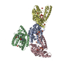
-Antibody , 2 types, 2 molecules LH
| #3: Antibody | Mass: 24086.316 Da / Num. of mol.: 1 Source method: isolated from a genetically manipulated source Source: (gene. exp.)  Homo sapiens (human) Homo sapiens (human) |
|---|---|
| #4: Antibody | Mass: 24222.092 Da / Num. of mol.: 1 Source method: isolated from a genetically manipulated source Source: (gene. exp.) Homo sapiens (human) |
-Sugars , 2 types, 3 molecules
| #5: Polysaccharide | alpha-D-mannopyranose-(1-3)-alpha-D-mannopyranose-(1-3)-beta-D-mannopyranose-(1-4)-2-acetamido-2- ...alpha-D-mannopyranose-(1-3)-alpha-D-mannopyranose-(1-3)-beta-D-mannopyranose-(1-4)-2-acetamido-2-deoxy-beta-D-glucopyranose-(1-4)-2-acetamido-2-deoxy-beta-D-glucopyranose Source method: isolated from a genetically manipulated source |
|---|---|
| #6: Polysaccharide | Source method: isolated from a genetically manipulated source |
-Details
| Has ligand of interest | N |
|---|---|
| Has protein modification | Y |
-Experimental details
-Experiment
| Experiment | Method: X-RAY DIFFRACTION / Number of used crystals: 1 |
|---|
- Sample preparation
| Crystal | Density Matthews: 6.16 Å3/Da / Density % sol: 80.02 % |
|---|---|
| Crystal grow | Temperature: 293 K / Method: vapor diffusion, hanging drop / Details: 20% PEG 400, CHES pH 9.0, 100mM MgSO4 |
-Data collection
| Diffraction | Mean temperature: 100 K / Serial crystal experiment: N |
|---|---|
| Diffraction source | Source: SYNCHROTRON / Site: ALS / Beamline: 8.2.2 / Wavelength: 0.999935 Å |
| Detector | Type: ADSC QUANTUM 315r / Detector: CCD / Date: Feb 26, 2015 |
| Radiation | Protocol: SINGLE WAVELENGTH / Monochromatic (M) / Laue (L): M / Scattering type: x-ray |
| Radiation wavelength | Wavelength: 0.999935 Å / Relative weight: 1 |
| Reflection | Resolution: 3.9→48.15 Å / Num. obs: 25174 / % possible obs: 97.65 % / Redundancy: 1.1 % / CC1/2: 1 / Net I/σ(I): 8.3 |
| Reflection shell | Resolution: 3.901→4.04 Å / Num. unique obs: 2324 / CC1/2: 1 |
-Phasing
| Phasing | Method: molecular replacement |
|---|
- Processing
| Software |
| ||||||||||||||||||||||||||||||||||||||||||||||||||||||||||||||||||||||
|---|---|---|---|---|---|---|---|---|---|---|---|---|---|---|---|---|---|---|---|---|---|---|---|---|---|---|---|---|---|---|---|---|---|---|---|---|---|---|---|---|---|---|---|---|---|---|---|---|---|---|---|---|---|---|---|---|---|---|---|---|---|---|---|---|---|---|---|---|---|---|---|
| Refinement | Method to determine structure: MOLECULAR REPLACEMENT Starting model: 5UGY Resolution: 3.901→48.15 Å / SU ML: 0.47 / Cross valid method: THROUGHOUT / σ(F): 1.35 / Phase error: 25.7 / Stereochemistry target values: ML
| ||||||||||||||||||||||||||||||||||||||||||||||||||||||||||||||||||||||
| Solvent computation | Shrinkage radii: 0.9 Å / VDW probe radii: 1.11 Å / Solvent model: FLAT BULK SOLVENT MODEL | ||||||||||||||||||||||||||||||||||||||||||||||||||||||||||||||||||||||
| Displacement parameters | Biso max: 259.36 Å2 / Biso mean: 91.99 Å2 / Biso min: 22.91 Å2 | ||||||||||||||||||||||||||||||||||||||||||||||||||||||||||||||||||||||
| Refinement step | Cycle: final / Resolution: 3.901→48.15 Å
| ||||||||||||||||||||||||||||||||||||||||||||||||||||||||||||||||||||||
| LS refinement shell | Refine-ID: X-RAY DIFFRACTION / Rfactor Rfree error: 0 / Total num. of bins used: 9
|
