 Movie
Movie Controller

[English] 日本語
 Yorodumi
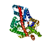
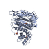
- PDB-1tae: Structural rearrangement accompanying NAD+ synthesis within a bac... -
+ Open data

- Basic information
| Entry | Database: PDB / ID: 1tae | ||||||
|---|---|---|---|---|---|---|---|
| Title | Structural rearrangement accompanying NAD+ synthesis within a bacterial DNA ligase crystal | ||||||
 Components Components | DNA ligase, NAD-dependent | ||||||
 Keywords Keywords | LIGASE / Nucleotidyl transferase fold | ||||||
| Function / homology |  Function and homology information Function and homology informationDNA ligase (NAD+) / DNA ligase (NAD+) activity / DNA replication / DNA repair / metal ion binding / cytosol Similarity search - Function | ||||||
| Biological species |   Enterococcus faecalis (bacteria) Enterococcus faecalis (bacteria) | ||||||
| Method |  X-RAY DIFFRACTION / MOLECULAR REPLACEMENT / Resolution: 2.7 Å X-RAY DIFFRACTION / MOLECULAR REPLACEMENT / Resolution: 2.7 Å | ||||||
 Authors Authors | Gajiwala, K.S. / Pinko, C. | ||||||
 Citation Citation | Journal: STRUCTURE / Year: 2004 Title: Structural rearrangement accompanying NAD+ synthesis within a bacterial DNA ligase crystal. Authors: Gajiwala, K.S. / Pinko, C. #1: Journal: Structure / Year: 1999Title: Structure of the adenylation domain of an NAD dependent DNA ligase Authors: Singleton, M.R. / Hakansson, K. / Timson, D.J. / Wigley, D.B. #2: Journal: Embo J. / Year: 2000Title: Crystal structure of NAD dependent DNA ligase: modular architecture and functional implications. Authors: Lee, J.Y. / Chang, C. / Song, H.K. / Moon, J. / Yang, J.K. / K Kim, H. / Kwon, S.T. / Suh, S.W. | ||||||
| History |
|
- Structure visualization
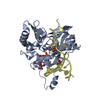
| Structure viewer | Molecule: MolmilJmol/JSmol |
|---|
- Downloads & links
-Download
| PDBx/mmCIF format | 1tae.cif.gz | 250.8 KB | Display | PDBx/mmCIF format |
|---|---|---|---|---|
| PDB format | pdb1tae.ent.gz | 206.5 KB | Display | PDB format |
| PDBx/mmJSON format | 1tae.json.gz | Tree view | PDBx/mmJSON format | |
| Others |  Other downloads Other downloads |
-Validation report
| Arichive directory | https://data.pdbj.org/pub/pdb/validation_reports/ta/1taeftp://data.pdbj.org/pub/pdb/validation_reports/ta/1tae | HTTPS FTP |
|---|
-Related structure data
| Related structure data | 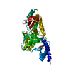 1ta8SC S: Starting model for refinement C: citing same article ( |
|---|---|
| Similar structure data |


-Links
 PDBj
PDBj


- Assembly
| Deposited unit | 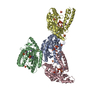
| ||||||||
|---|---|---|---|---|---|---|---|---|---|
| 1 | 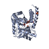
| ||||||||
| 2 | 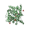
| ||||||||
| 3 | 
| ||||||||
| 4 | 
| ||||||||
| 5 | 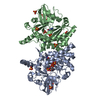
| ||||||||
| 6 | 
| ||||||||
| Unit cell |
|
-Components
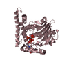
| #1: Protein | Mass: 38032.688 Da / Num. of mol.: 4 / Fragment: Adenylation domain Source method: isolated from a genetically manipulated source Source: (gene. exp.) Enterococcus faecalis (bacteria) / Strain: V583 / Gene: ligA / Production host: References: UniProt: Q837V6, GenBank: 29375318, DNA ligase (NAD+) #2: Chemical | ChemComp-SO4 /   Mass: 96.063 Da / Num. of mol.: 24 / Source method: obtained synthetically / Formula: SO4 Mass: 96.063 Da / Num. of mol.: 24 / Source method: obtained synthetically / Formula: SO4#3: Chemical | ChemComp-NAD /   Mass: 663.425 Da / Num. of mol.: 4 / Source method: obtained synthetically / Formula: C21H27N7O14P2 / Comment: NAD*YM Mass: 663.425 Da / Num. of mol.: 4 / Source method: obtained synthetically / Formula: C21H27N7O14P2 / Comment: NAD*YM#4: Chemical |   Mass: 22.990 Da / Num. of mol.: 2 / Source method: obtained synthetically / Formula: Na Mass: 22.990 Da / Num. of mol.: 2 / Source method: obtained synthetically / Formula: Na#5: Water | ChemComp-HOH / |  Mass: 18.015 Da / Num. of mol.: 176 / Source method: isolated from a natural source / Formula: H2O Mass: 18.015 Da / Num. of mol.: 176 / Source method: isolated from a natural source / Formula: H2O |
|---|
-Experimental details
-Experiment
| Experiment | Method: X-RAY DIFFRACTION / Number of used crystals: 1 |
|---|
- Sample preparation
| Crystal | Density Matthews: 3.13 Å3/Da / Density % sol: 60.7 % |
|---|---|
| Crystal grow | Temperature: 286 K / Method: vapor diffusion, hanging drop / pH: 5.5 Details: Ammonium sulfate, cacodylate, pH 5.5, VAPOR DIFFUSION, HANGING DROP, temperature 286K |
-Data collection
| Diffraction | Mean temperature: 100 K |
|---|---|
| Diffraction source | Source: ROTATING ANODE / Type: ENRAF-NONIUS FR591 / Wavelength: 1.5418 Å |
| Detector | Type: MARRESEARCH / Detector: IMAGE PLATE / Date: Jan 9, 2004 / Details: Osmic Confocal Max-Flux Optics |
| Radiation | Monochromator: Osmic mirrors / Protocol: SINGLE WAVELENGTH / Monochromatic (M) / Laue (L): M / Scattering type: x-ray |
| Radiation wavelength | Wavelength: 1.5418 Å / Relative weight: 1 |
| Reflection | Resolution: 2.7→25 Å / Num. all: 51184 / Num. obs: 50300 / % possible obs: 98.3 % / Observed criterion σ(F): 1 / Observed criterion σ(I): 1 / Redundancy: 3.84 % / Biso Wilson estimate: 56.4 Å2 / Rsym value: 0.074 / Net I/σ(I): 13.1 |
| Reflection shell | Resolution: 2.7→2.8 Å / Redundancy: 3.6 % / Mean I/σ(I) obs: 4.2 / Num. unique all: 4916 / Rsym value: 0.374 / % possible all: 97.3 |
- Processing
| Software |
| |||||||||||||||||||||||||
|---|---|---|---|---|---|---|---|---|---|---|---|---|---|---|---|---|---|---|---|---|---|---|---|---|---|---|
| Refinement | Method to determine structure: MOLECULAR REPLACEMENT Starting model: Domain 1b of NMN bound structure of E. faecalis ligase (PDB ID: 1TA8) Resolution: 2.7→24.77 Å / Rfactor Rfree error: 0.004 / Data cutoff high absF: 482888.09 / Data cutoff low absF: 0 / Isotropic thermal model: RESTRAINED / Cross valid method: THROUGHOUT / σ(F): 0 / Stereochemistry target values: Engh & Huber
| |||||||||||||||||||||||||
| Solvent computation | Solvent model: FLAT MODEL / Bsol: 44.2795 Å2 / ksol: 0.369729 e/Å3 | |||||||||||||||||||||||||
| Displacement parameters | Biso mean: 43.8 Å2
| |||||||||||||||||||||||||
| Refine analyze |
| |||||||||||||||||||||||||
| Refinement step | Cycle: LAST / Resolution: 2.7→24.77 Å
| |||||||||||||||||||||||||
| Refine LS restraints |
| |||||||||||||||||||||||||
| LS refinement shell | Resolution: 2.7→2.87 Å / Rfactor Rfree error: 0.014 / Total num. of bins used: 6
| |||||||||||||||||||||||||
| Xplor file |
|
