 Movie
Movie Controller


+ Open data

- Basic information
| Entry | Database: PDB / ID: 7jgz | ||||||
|---|---|---|---|---|---|---|---|
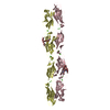
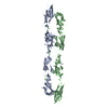
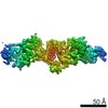
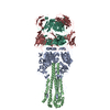
| Title | Protocadherin gammaC4 EC1-4 crystal structure | ||||||
 Components Components | Protocadherin gamma C4 | ||||||
 Keywords Keywords | CELL ADHESION / Protocadherin / Cadherin / Cell-surface receptor / Neuronal self-avoidance | ||||||
| Function / homology |  Function and homology information Function and homology informationhomophilic cell adhesion via plasma membrane adhesion molecules / synapse organization / negative regulation of neuron apoptotic process / calcium ion binding / membrane / plasma membrane Similarity search - Function | ||||||
| Biological species |  | ||||||
| Method |  X-RAY DIFFRACTION / SYNCHROTRON / MOLECULAR REPLACEMENT / Resolution: 3.51 Å X-RAY DIFFRACTION / SYNCHROTRON / MOLECULAR REPLACEMENT / Resolution: 3.51 Å | ||||||
 Authors Authors | Goodman, K.M. / Mannepalli, S. / Honig, B. / Shapiro, L. | ||||||
| Funding support |  United States, 1items United States, 1items
| ||||||
 Citation Citation | Journal: Elife / Year: 2022 Title: How clustered protocadherin binding specificity is tuned for neuronal self-/nonself-recognition. Authors: Goodman, K.M. / Katsamba, P.S. / Rubinstein, R. / Ahlsen, G. / Bahna, F. / Mannepalli, S. / Dan, H. / Sampogna, R.V. / Shapiro, L. / Honig, B. | ||||||
| History |
|
- Structure visualization
| Structure viewer | Molecule: MolmilJmol/JSmol |
|---|
- Downloads & links
-Download
| PDBx/mmCIF format | 7jgz.cif.gz | 214.6 KB | Display | PDBx/mmCIF format |
|---|---|---|---|---|
| PDB format | pdb7jgz.ent.gz | 146 KB | Display | PDB format |
| PDBx/mmJSON format | 7jgz.json.gz | Tree view | PDBx/mmJSON format | |
| Others |  Other downloads Other downloads |
-Validation report
| Summary document | 7jgz_validation.pdf.gz | 1 MB | Display | wwPDB validaton report |
|---|---|---|---|---|
| Full document | 7jgz_full_validation.pdf.gz | 1 MB | Display | |
| Data in XML | 7jgz_validation.xml.gz | 17 KB | Display | |
| Data in CIF | 7jgz_validation.cif.gz | 22.5 KB | Display | |
| Arichive directory | https://data.pdbj.org/pub/pdb/validation_reports/jg/7jgzftp://data.pdbj.org/pub/pdb/validation_reports/jg/7jgz | HTTPS FTP |
-Related structure data
| Related structure data |  7rgfC 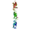 4zpoS  6wzh S: Starting model for refinement C: citing same article ( |
|---|---|
| Similar structure data |


-Links
 PDBj
PDBj


- Assembly
| Deposited unit | 
| ||||||||||
|---|---|---|---|---|---|---|---|---|---|---|---|
| 1 | 
| ||||||||||
| Unit cell |
|
-Components
-Protein , 1 types, 1 molecules A
| #1: Protein | Mass: 47006.918 Da / Num. of mol.: 1 Source method: isolated from a genetically manipulated source Source: (gene. exp.)  Homo sapiens (human) / References: UniProt: Q91XX0 Homo sapiens (human) / References: UniProt: Q91XX0 |
|---|
-Sugars , 3 types, 4 molecules 
| #2: Polysaccharide | beta-D-mannopyranose-(1-3)-2-acetamido-2-deoxy-beta-D-glucopyranose-(1-4)-[alpha-L-fucopyranose-(1- ...beta-D-mannopyranose-(1-3)-2-acetamido-2-deoxy-beta-D-glucopyranose-(1-4)-[alpha-L-fucopyranose-(1-6)]2-acetamido-2-deoxy-beta-D-glucopyranose Source method: isolated from a genetically manipulated source |
|---|---|
| #3: Polysaccharide | 2-acetamido-2-deoxy-beta-D-glucopyranose-(1-4)-[alpha-L-fucopyranose-(1-6)]2-acetamido-2-deoxy-beta- ...2-acetamido-2-deoxy-beta-D-glucopyranose-(1-4)-[alpha-L-fucopyranose-(1-6)]2-acetamido-2-deoxy-beta-D-glucopyranose Source method: isolated from a genetically manipulated source |
| #4: Sugar |  Type: D-saccharide, alpha linking / Mass: 180.156 Da / Num. of mol.: 2 / Source method: obtained synthetically / Formula: C6H12O6 Type: D-saccharide, alpha linking / Mass: 180.156 Da / Num. of mol.: 2 / Source method: obtained synthetically / Formula: C6H12O6 |
-Non-polymers , 2 types, 10 molecules 

| #5: Chemical | ChemComp-CA /  Mass: 40.078 Da / Num. of mol.: 9 Mass: 40.078 Da / Num. of mol.: 9Source method: isolated from a genetically manipulated source Formula: Ca #6: Chemical | ChemComp-EDO / |  Mass: 62.068 Da / Num. of mol.: 1 Mass: 62.068 Da / Num. of mol.: 1Source method: isolated from a genetically manipulated source Formula: C2H6O2 |
|---|
-Details
| Has ligand of interest | N |
|---|---|
| Has protein modification | Y |
-Experimental details
-Experiment
| Experiment | Method: X-RAY DIFFRACTION / Number of used crystals: 1 |
|---|
- Sample preparation
| Crystal | Density Matthews: 4.5 Å3/Da / Density % sol: 72.67 % |
|---|---|
| Crystal grow | Temperature: 293 K / Method: batch mode Details: 10% (w/v) PEG8000, 20% ethylene glycol, 10% Morpheus Amino Acids (Molecular Dimensions), and 0.1 M Morpheus Buffer System 2 (Hepes/MOPS buffer) pH 7.5 |
-Data collection
| Diffraction | Mean temperature: 100 K / Serial crystal experiment: N |
|---|---|
| Diffraction source | Source: SYNCHROTRON / Site: APS / Beamline: 24-ID-E / Wavelength: 0.97918 Å |
| Detector | Type: ADSC QUANTUM 315 / Detector: CCD / Date: Feb 7, 2017 |
| Radiation | Protocol: SINGLE WAVELENGTH / Monochromatic (M) / Laue (L): M / Scattering type: x-ray |
| Radiation wavelength | Wavelength: 0.97918 Å / Relative weight: 1 |
| Reflection | Resolution: 3.5→40 Å / Num. obs: 7545 / % possible obs: 67.6 % / Redundancy: 7.1 % / Biso Wilson estimate: 77.57 Å2 / CC1/2: 0.997 / Rmerge(I) obs: 0.215 / Rpim(I) all: 0.086 / Rrim(I) all: 0.232 / Net I/σ(I): 8.3 |
| Reflection shell | Resolution: 3.5→3.84 Å / Redundancy: 7.1 % / Rmerge(I) obs: 0.83 / Mean I/σ(I) obs: 2.9 / Num. unique obs: 340 / CC1/2: 0.829 / Rpim(I) all: 0.335 / Rrim(I) all: 0.896 / % possible all: 12.5 |
- Processing
| Software |
| |||||||||||||||||||||||||||||||||||||||||||||||||||||||||||||||||||||||||||||||||||||||||||||||||||||||||||||||||||||||||||||
|---|---|---|---|---|---|---|---|---|---|---|---|---|---|---|---|---|---|---|---|---|---|---|---|---|---|---|---|---|---|---|---|---|---|---|---|---|---|---|---|---|---|---|---|---|---|---|---|---|---|---|---|---|---|---|---|---|---|---|---|---|---|---|---|---|---|---|---|---|---|---|---|---|---|---|---|---|---|---|---|---|---|---|---|---|---|---|---|---|---|---|---|---|---|---|---|---|---|---|---|---|---|---|---|---|---|---|---|---|---|---|---|---|---|---|---|---|---|---|---|---|---|---|---|---|---|---|
| Refinement | Method to determine structure: MOLECULAR REPLACEMENT Starting model: 4ZPO Resolution: 3.51→38.5 Å / SU ML: 0.4166 / Cross valid method: THROUGHOUT / σ(F): 1.36 / Phase error: 25.8692 / Stereochemistry target values: GeoStd + Monomer Library Details: Please note ellipsoidal resolution limits of 4.6/3.9/3.5 were applied to the data. The completeness within the ellipsoidal limits is 96.8%.
| |||||||||||||||||||||||||||||||||||||||||||||||||||||||||||||||||||||||||||||||||||||||||||||||||||||||||||||||||||||||||||||
| Solvent computation | Shrinkage radii: 0.9 Å / VDW probe radii: 1.11 Å / Solvent model: FLAT BULK SOLVENT MODEL | |||||||||||||||||||||||||||||||||||||||||||||||||||||||||||||||||||||||||||||||||||||||||||||||||||||||||||||||||||||||||||||
| Displacement parameters | Biso mean: 101.24 Å2 | |||||||||||||||||||||||||||||||||||||||||||||||||||||||||||||||||||||||||||||||||||||||||||||||||||||||||||||||||||||||||||||
| Refinement step | Cycle: LAST / Resolution: 3.51→38.5 Å
| |||||||||||||||||||||||||||||||||||||||||||||||||||||||||||||||||||||||||||||||||||||||||||||||||||||||||||||||||||||||||||||
| Refine LS restraints |
| |||||||||||||||||||||||||||||||||||||||||||||||||||||||||||||||||||||||||||||||||||||||||||||||||||||||||||||||||||||||||||||
| LS refinement shell |
| |||||||||||||||||||||||||||||||||||||||||||||||||||||||||||||||||||||||||||||||||||||||||||||||||||||||||||||||||||||||||||||
| Refinement TLS params. | Method: refined / Refine-ID: X-RAY DIFFRACTION
| |||||||||||||||||||||||||||||||||||||||||||||||||||||||||||||||||||||||||||||||||||||||||||||||||||||||||||||||||||||||||||||
| Refinement TLS group |
|
