 Movie
Movie Controller

[English] 日本語
 Yorodumi
- PDB-7jfs: The structure of the CBM32-1, CBM32-2, and M60 catalytic domains ... -
+ Open data

- Basic information
| Entry | Database: PDB / ID: 7jfs | |||||||||
|---|---|---|---|---|---|---|---|---|---|---|
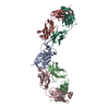
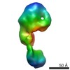
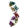
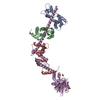
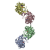
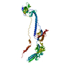
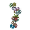
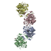
| Title | The structure of the CBM32-1, CBM32-2, and M60 catalytic domains from Clostridium perfringens ZmpB | |||||||||
 Components Components | F5/8 type C domain protein | |||||||||
 Keywords Keywords | HYDROLASE / glycopeptidase | |||||||||
| Function / homology |  Function and homology information Function and homology information | |||||||||
| Biological species |   Clostridium perfringens (bacteria) Clostridium perfringens (bacteria) | |||||||||
| Method |  X-RAY DIFFRACTION / MOLECULAR REPLACEMENT / molecular replacement / Resolution: 4.6 Å X-RAY DIFFRACTION / MOLECULAR REPLACEMENT / molecular replacement / Resolution: 4.6 Å | |||||||||
 Authors Authors | Pluvinage, B. / Boraston, A.B. | |||||||||
| Funding support |  Canada, 2items Canada, 2items
| |||||||||
 Citation Citation | Journal: Proc.Natl.Acad.Sci.USA / Year: 2021 Title: Architecturally complex O -glycopeptidases are customized for mucin recognition and hydrolysis. Authors: Pluvinage, B. / Ficko-Blean, E. / Noach, I. / Stuart, C. / Thompson, N. / McClure, H. / Buenbrazo, N. / Wakarchuk, W. / Boraston, A.B. | |||||||||
| History |
|
- Structure visualization
| Structure viewer | Molecule: MolmilJmol/JSmol |
|---|
- Downloads & links
-Download
| PDBx/mmCIF format | 7jfs.cif.gz | 205.7 KB | Display | PDBx/mmCIF format |
|---|---|---|---|---|
| PDB format | pdb7jfs.ent.gz | 158.8 KB | Display | PDB format |
| PDBx/mmJSON format | 7jfs.json.gz | Tree view | PDBx/mmJSON format | |
| Others |  Other downloads Other downloads |
-Validation report
| Arichive directory | https://data.pdbj.org/pub/pdb/validation_reports/jf/7jfsftp://data.pdbj.org/pub/pdb/validation_reports/jf/7jfs | HTTPS FTP |
|---|
-Related structure data
| Related structure data | 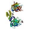 7jnbC 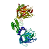 7jndC 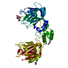 7jnfC 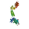 7jrlC 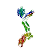 7jrmC 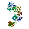 7js4C 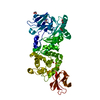 5kdnS S: Starting model for refinement C: citing same article ( |
|---|---|
| Similar structure data |
-Links
 PDBj
PDBj


- Assembly
| Deposited unit | 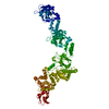
| ||||||||
|---|---|---|---|---|---|---|---|---|---|
| 1 |
| ||||||||
| Unit cell |
|
-Components
| #1: Protein | Mass: 112008.195 Da / Num. of mol.: 1 / Fragment: UNP residues 46-1003 Source method: isolated from a genetically manipulated source Source: (gene. exp.) Clostridium perfringens (strain ATCC 13124 / DSM 756 / JCM 1290 / NCIMB 6125 / NCTC 8237 / Type A) (bacteria)Strain: ATCC 13124 / DSM 756 / JCM 1290 / NCIMB 6125 / NCTC 8237 / Type A Gene: CPF_1489 / Plasmid: pET28a / Production host: |
|---|---|
| #2: Chemical | ChemComp-ZN /   Mass: 65.409 Da / Num. of mol.: 1 / Source method: obtained synthetically / Formula: Zn Mass: 65.409 Da / Num. of mol.: 1 / Source method: obtained synthetically / Formula: Zn |
| Has ligand of interest | N |
-Experimental details
-Experiment
| Experiment | Method: X-RAY DIFFRACTION / Number of used crystals: 1 |
|---|
- Sample preparation
| Crystal | Density Matthews: 3.63 Å3/Da / Density % sol: 66.09 % |
|---|---|
| Crystal grow | Temperature: 291 K / Method: vapor diffusion, hanging drop / pH: 6 / Details: 2% Tascimate, 0.1 M Bis-Tris, 15% PEG3350 |
-Data collection
| Diffraction | Mean temperature: 100 K / Serial crystal experiment: N |
|---|---|
| Diffraction source | Source: ROTATING ANODE / Type: RIGAKU MICROMAX-002 / Wavelength: 1.54187 Å |
| Detector | Type: DECTRIS PILATUS 200K / Detector: PIXEL / Date: Feb 14, 2020 |
| Radiation | Protocol: SINGLE WAVELENGTH / Monochromatic (M) / Laue (L): M / Scattering type: x-ray |
| Radiation wavelength | Wavelength: 1.54187 Å / Relative weight: 1 |
| Reflection | Resolution: 4.6→30 Å / Num. obs: 9041 / % possible obs: 99.7 % / Redundancy: 4.7 % / CC1/2: 0.981 / Rmerge(I) obs: 0.183 / Rpim(I) all: 0.084 / Net I/σ(I): 8.6 |
| Reflection shell | Resolution: 4.6→4.68 Å / Redundancy: 4.4 % / Rmerge(I) obs: 0.757 / Mean I/σ(I) obs: 1.9 / Num. unique obs: 449 / CC1/2: 0.798 / Rpim(I) all: 0.387 / % possible all: 100 |
-Phasing
| Phasing | Method: molecular replacement |
|---|
- Processing
| Software |
| ||||||||||||||||||||||||||||
|---|---|---|---|---|---|---|---|---|---|---|---|---|---|---|---|---|---|---|---|---|---|---|---|---|---|---|---|---|---|
| Refinement | Method to determine structure: MOLECULAR REPLACEMENT Starting model: PDB entry 5KDN Resolution: 4.6→29.93 Å / SU ML: 0.65 / Cross valid method: THROUGHOUT / σ(F): 1.35 / Phase error: 32.93 / Stereochemistry target values: ML
| ||||||||||||||||||||||||||||
| Solvent computation | Shrinkage radii: 0.9 Å / VDW probe radii: 1.11 Å / Solvent model: FLAT BULK SOLVENT MODEL | ||||||||||||||||||||||||||||
| Displacement parameters | Biso max: 219.36 Å2 / Biso mean: 96.0881 Å2 / Biso min: 59.42 Å2 | ||||||||||||||||||||||||||||
| Refinement step | Cycle: final / Resolution: 4.6→29.93 Å
| ||||||||||||||||||||||||||||
| LS refinement shell | Refine-ID: X-RAY DIFFRACTION / Rfactor Rfree error: 0 / Total num. of bins used: 3
|
