 Movie
Movie Controller

[English] 日本語
 Yorodumi
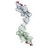
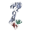
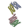
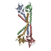
- PDB-7jrl: The structure of CBM51-2 in complex with GlcNAc and INT domains f... -
+ Open data

- Basic information
| Entry | Database: PDB / ID: 7jrl | |||||||||
|---|---|---|---|---|---|---|---|---|---|---|
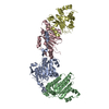
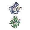
| Title | The structure of CBM51-2 in complex with GlcNAc and INT domains from Clostridium perfringens ZmpB | |||||||||
 Components Components | F5/8 type C domain protein | |||||||||
 Keywords Keywords | SUGAR BINDING PROTEIN / Carbohydrate binding module | |||||||||
| Function / homology |  Function and homology information Function and homology information | |||||||||
| Biological species |   Clostridium perfringens (bacteria) Clostridium perfringens (bacteria) | |||||||||
| Method |  X-RAY DIFFRACTION / SYNCHROTRON / MOLECULAR REPLACEMENT / molecular replacement / Resolution: 1.5 Å X-RAY DIFFRACTION / SYNCHROTRON / MOLECULAR REPLACEMENT / molecular replacement / Resolution: 1.5 Å | |||||||||
 Authors Authors | Pluvinage, B. / Boraston, A.B. | |||||||||
| Funding support |  Canada, 2items Canada, 2items
| |||||||||
 Citation Citation | Journal: Proc.Natl.Acad.Sci.USA / Year: 2021 Title: Architecturally complex O -glycopeptidases are customized for mucin recognition and hydrolysis. Authors: Pluvinage, B. / Ficko-Blean, E. / Noach, I. / Stuart, C. / Thompson, N. / McClure, H. / Buenbrazo, N. / Wakarchuk, W. / Boraston, A.B. | |||||||||
| History |
|
- Structure visualization
| Structure viewer | Molecule: MolmilJmol/JSmol |
|---|
- Downloads & links
-Download
| PDBx/mmCIF format | 7jrl.cif.gz | 120.7 KB | Display | PDBx/mmCIF format |
|---|---|---|---|---|
| PDB format | pdb7jrl.ent.gz | 88.3 KB | Display | PDB format |
| PDBx/mmJSON format | 7jrl.json.gz | Tree view | PDBx/mmJSON format | |
| Others |  Other downloads Other downloads |
-Validation report
| Arichive directory | https://data.pdbj.org/pub/pdb/validation_reports/jr/7jrlftp://data.pdbj.org/pub/pdb/validation_reports/jr/7jrl | HTTPS FTP |
|---|
-Related structure data
| Related structure data | 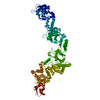 7jfsC 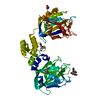 7jnbC 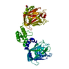 7jndC 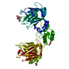 7jnfC 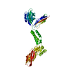 7jrmC 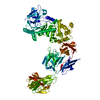 7js4C  2vmgS S: Starting model for refinement C: citing same article ( |
|---|---|
| Similar structure data |


-Links
 PDBj
PDBj


- Assembly
| Deposited unit | 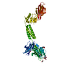
| ||||||||
|---|---|---|---|---|---|---|---|---|---|
| 1 |
| ||||||||
| Unit cell |
|
-Components
-Protein / Sugars , 2 types, 2 molecules A
| #1: Protein | Mass: 53592.801 Da / Num. of mol.: 1 Fragment: Carbohydrate binding module (UNP residues 1227-1687) Source method: isolated from a genetically manipulated source Source: (gene. exp.) Clostridium perfringens (strain ATCC 13124 / DSM 756 / JCM 1290 / NCIMB 6125 / NCTC 8237 / Type A) (bacteria)Strain: ATCC 13124 / DSM 756 / JCM 1290 / NCIMB 6125 / NCTC 8237 / Type A Gene: CPF_1489 / Plasmid: pET28a / Production host: |
|---|---|
| #3: Sugar | ChemComp-NAG /  Type: D-saccharide, beta linking / Mass: 221.208 Da / Num. of mol.: 1 / Source method: obtained synthetically / Formula: C8H15NO6 / Feature type: SUBJECT OF INVESTIGATION Type: D-saccharide, beta linking / Mass: 221.208 Da / Num. of mol.: 1 / Source method: obtained synthetically / Formula: C8H15NO6 / Feature type: SUBJECT OF INVESTIGATION |
-Non-polymers , 5 types, 554 molecules 


| #2: Chemical | ChemComp-CA /  Mass: 40.078 Da / Num. of mol.: 1 / Source method: obtained synthetically / Formula: Ca Mass: 40.078 Da / Num. of mol.: 1 / Source method: obtained synthetically / Formula: Ca | ||||||
|---|---|---|---|---|---|---|---|
| #4: Chemical | ChemComp-EDO /  Mass: 62.068 Da / Num. of mol.: 4 / Source method: obtained synthetically / Formula: C2H6O2 Mass: 62.068 Da / Num. of mol.: 4 / Source method: obtained synthetically / Formula: C2H6O2#5: Chemical | ChemComp-PO4 / |  Mass: 94.971 Da / Num. of mol.: 1 / Source method: obtained synthetically / Formula: PO4 Mass: 94.971 Da / Num. of mol.: 1 / Source method: obtained synthetically / Formula: PO4#6: Chemical | ChemComp-TRS / |  Mass: 122.143 Da / Num. of mol.: 1 / Source method: obtained synthetically / Formula: C4H12NO3 / Comment: pH buffer*YM Mass: 122.143 Da / Num. of mol.: 1 / Source method: obtained synthetically / Formula: C4H12NO3 / Comment: pH buffer*YM#7: Water | ChemComp-HOH / | Mass: 18.015 Da / Num. of mol.: 547 / Source method: isolated from a natural source / Formula: H2O |
-Details
| Has ligand of interest | Y |
|---|
-Experimental details
-Experiment
| Experiment | Method: X-RAY DIFFRACTION / Number of used crystals: 1 |
|---|
- Sample preparation
| Crystal | Density Matthews: 2.54 Å3/Da / Density % sol: 51.55 % |
|---|---|
| Crystal grow | Temperature: 291 K / Method: vapor diffusion, hanging drop / pH: 7 Details: 0.15 M sodium phosphate monobasic, 18% PEG3350, 0.1 M Tris-HCl |
-Data collection
| Diffraction | Mean temperature: 100 K / Serial crystal experiment: N |
|---|---|
| Diffraction source | Source: SYNCHROTRON / Site: SSRL  / Beamline: BL9-2 / Wavelength: 0.91966 Å / Beamline: BL9-2 / Wavelength: 0.91966 Å |
| Detector | Type: MARMOSAIC 325 mm CCD / Detector: CCD / Date: Apr 11, 2011 |
| Radiation | Monochromator: Liquid nitrogen-cooled double crystal Si(111) Protocol: SINGLE WAVELENGTH / Monochromatic (M) / Laue (L): M / Scattering type: x-ray |
| Radiation wavelength | Wavelength: 0.91966 Å / Relative weight: 1 |
| Reflection | Resolution: 1.5→42.86 Å / Num. obs: 86269 / % possible obs: 100 % / Redundancy: 5 % / CC1/2: 0.998 / Rmerge(I) obs: 0.082 / Rpim(I) all: 0.04 / Net I/σ(I): 13.2 |
| Reflection shell | Resolution: 1.5→1.58 Å / Redundancy: 5 % / Rmerge(I) obs: 0.581 / Mean I/σ(I) obs: 3.6 / Num. unique obs: 12539 / CC1/2: 0.735 / Rpim(I) all: 0.283 / % possible all: 100 |
-Phasing
| Phasing | Method: molecular replacement |
|---|
- Processing
| Software |
| |||||||||||||||||||||||||||||||||||||||||||||
|---|---|---|---|---|---|---|---|---|---|---|---|---|---|---|---|---|---|---|---|---|---|---|---|---|---|---|---|---|---|---|---|---|---|---|---|---|---|---|---|---|---|---|---|---|---|---|
| Refinement | Method to determine structure: MOLECULAR REPLACEMENT Starting model: PDB entry 2VMG Resolution: 1.5→38.93 Å / Cor.coef. Fo:Fc: 0.955 / Cor.coef. Fo:Fc free: 0.951 / SU B: 1.147 / SU ML: 0.043 / Cross valid method: THROUGHOUT / σ(F): 0 / ESU R: 0.071 / ESU R Free: 0.067 / Stereochemistry target values: MAXIMUM LIKELIHOOD / Details: U VALUES : REFINED INDIVIDUALLY
| |||||||||||||||||||||||||||||||||||||||||||||
| Solvent computation | Ion probe radii: 0.8 Å / Shrinkage radii: 0.8 Å / VDW probe radii: 1.2 Å / Solvent model: MASK | |||||||||||||||||||||||||||||||||||||||||||||
| Displacement parameters | Biso max: 62.74 Å2 / Biso mean: 16.24 Å2 / Biso min: 6.44 Å2
| |||||||||||||||||||||||||||||||||||||||||||||
| Refinement step | Cycle: final / Resolution: 1.5→38.93 Å
| |||||||||||||||||||||||||||||||||||||||||||||
| Refine LS restraints |
| |||||||||||||||||||||||||||||||||||||||||||||
| LS refinement shell | Resolution: 1.5→1.539 Å / Rfactor Rfree error: 0 / Total num. of bins used: 20
|
