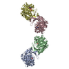
- PDB-4r8g: Crystal Structure of Myosin-1c tail in complex with Calmodulin -
+
Open data
ID or keywords:
Loading...
-
Basic information
Entry
Database: PDB / ID: 4r8g
Title
Crystal Structure of Myosin-1c tail in complex with Calmodulin
Components
Calmodulin
Unconventional myosin-Ic
Keywords
PROTEIN BINDING/CALCIUM-BINDING PROTEIN / EF hand / PH domain / IQ motif / Myosin / Ca2+ signaling / Force sensing / Calcium binding / lipid binding / plasma membrane / cytoskeleton / PROTEIN BINDING-CALCIUM-BINDING PROTEIN complex
Function / homology
Function and homology information
stereocilium membrane / B-WICH complex positively regulates rRNA expression / positive regulation of cellular response to insulin stimulus / vesicle transport along actin filament / Regulation of actin dynamics for phagocytic cup formation / stereocilium bundle / actin filament-based movement / B-WICH complex / stereocilium / myosin complex ...stereocilium membrane / B-WICH complex positively regulates rRNA expression / positive regulation of cellular response to insulin stimulus / vesicle transport along actin filament / Regulation of actin dynamics for phagocytic cup formation / stereocilium bundle / actin filament-based movement / B-WICH complex / stereocilium / myosin complex / myosin II complex / protein targeting to membrane / positive regulation of transcription by RNA polymerase III / regulation of bicellular tight junction assembly / vascular endothelial growth factor signaling pathway / microfilament motor activity / positive regulation of transcription by RNA polymerase I / filamentous actin / microvillus / positive regulation of actin filament polymerization / brush border / lateral plasma membrane / positive regulation of protein targeting to membrane / phagocytic vesicle / basal plasma membrane / cytoplasmic vesicle membrane / actin filament organization / phospholipid binding / cellular response to type II interferon / small GTPase binding / ruffle membrane / endocytosis / actin filament binding / actin cytoskeleton / actin binding / cell cortex / calmodulin binding / nuclear body / positive regulation of cell migration / chromatin remodeling / membrane raft / signaling receptor binding / calcium ion binding / nucleolus / positive regulation of transcription by RNA polymerase II / ATP binding / membrane / plasma membrane / cytoplasm / cytosol Similarity search - Function
Class I myosin tail homology domain / Class I myosin, motor domain / Unconventional myosin tail, actin- and lipid-binding / Class I myosin tail homology (TH1) domain profile. / IQ calmodulin-binding motif / Short calmodulin-binding motif containing conserved Ile and Gln residues. / IQ motif, EF-hand binding site / Myosin motor domain profile. / Myosin head, motor domain / Myosin head (motor domain) ...Class I myosin tail homology domain / Class I myosin, motor domain / Unconventional myosin tail, actin- and lipid-binding / Class I myosin tail homology (TH1) domain profile. / IQ calmodulin-binding motif / Short calmodulin-binding motif containing conserved Ile and Gln residues. / IQ motif, EF-hand binding site / Myosin motor domain profile. / Myosin head, motor domain / Myosin head (motor domain) / Myosin. Large ATPases. / IQ motif profile. / Kinesin motor domain superfamily / : / EF-hand domain pair / EF-hand, calcium binding motif / EF-Hand 1, calcium-binding site / EF-hand calcium-binding domain. / EF-hand calcium-binding domain profile. / EF-hand domain / EF-hand domain pair / P-loop containing nucleoside triphosphate hydrolase Similarity search - Domain/homology
In the structure databanks used in Yorodumi, some data are registered as the other names, "COVID-19 virus" and "2019-nCoV". Here are the details of the virus and the list of structure data.
Jan 31, 2019. EMDB accession codes are about to change! (news from PDBe EMDB page)
EMDB accession codes are about to change! (news from PDBe EMDB page)
The allocation of 4 digits for EMDB accession codes will soon come to an end. Whilst these codes will remain in use, new EMDB accession codes will include an additional digit and will expand incrementally as the available range of codes is exhausted. The current 4-digit format prefixed with “EMD-” (i.e. EMD-XXXX) will advance to a 5-digit format (i.e. EMD-XXXXX), and so on. It is currently estimated that the 4-digit codes will be depleted around Spring 2019, at which point the 5-digit format will come into force.
The EM Navigator/Yorodumi systems omit the EMD- prefix.
Related info.:Q: What is EMD? / ID/Accession-code notation in Yorodumi/EM Navigator
Yorodumi is a browser for structure data from EMDB, PDB, SASBDB, etc.
This page is also the successor to EM Navigator detail page, and also detail information page/front-end page for Omokage search.
The word "yorodu" (or yorozu) is an old Japanese word meaning "ten thousand". "mi" (miru) is to see.
Related info.:EMDB / PDB / SASBDB / Comparison of 3 databanks / Yorodumi Search / Aug 31, 2016. New EM Navigator & Yorodumi / Yorodumi Papers / Jmol/JSmol / Function and homology information / Changes in new EM Navigator and Yorodumi
 Movie
Movie Controller


 Open data
 Basic information
 Components
 Keywords
 Function and homology information
 X-RAY DIFFRACTION /
  Authors
 Citation
 Structure visualization
 Downloads & links
 Other downloads
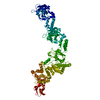


 PDBj
PDBj


 Assembly
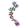


 Mass: 96.063 Da / Num. of mol.: 3 / Source method: obtained synthetically / Formula: SO4
 Sample preparation
 / Beamline: BL17U / Wavelength: 0.9793 Å
 Processing